Article Text

Abstract
Introduction: The 22q13.3 deletion syndrome (MIM 606232) is characterised by neonatal hypotonia, normal to accelerated growth, absent to severely delayed speech, global developmental delay, and minor dysmorphic facial features. We report the molecular characterisation of the deletion breakpoint in two unrelated chromosome 22q13.3 deletion cases.
Methods: The deletions were characterised by FISH, checked for other abnormalities by array-CGH, and confirmed by Real-Time PCR, and finally the breakpoints were cloned, sequenced, and compared.
Results: Both cases show the cardinal features of the 22q13.3 deletion syndrome associated with a deletion involving the last 100 kb of chromosome 22q13.3. The cases show a breakpoint within the same 15 bp repeat unit, overlapping the results obtained by Wong and colleagues in 1997 and suggesting that a recurrent deletion breakpoint exists within the SHANK3 gene. The direct repeat involved in these 22q13 deletion cases is presumably able to form slipped (hairpin) structures, but it also has a strong potential for forming tetraplex structures.
Discussion: Three cases with a common breakpoint within SHANK3 share a number of common phenotypic features, such as mental retardation and developmental delay with severely delayed or absent expressive speech. The two cases presented here, having a deletion partially overlapping the commercial subtelomeric probe, highlight the difficulties in interpreting FISH results and suggest that many similar cases may be overlooked.
- AE, age equivalent
- PEP-R, Psycho-Educational Profile-Revised
- PSD, post-synaptic density
- VABS, Vineland Adaptative Behaviour Scale
- 22q13 deletion syndrome
- FISH
- recurrent deletion
- SHANK3
- subtelomeric deletion
Statistics from Altmetric.com
- AE, age equivalent
- PEP-R, Psycho-Educational Profile-Revised
- PSD, post-synaptic density
- VABS, Vineland Adaptative Behaviour Scale
Cryptic deletions involving the distal portions of chromosomes have been demonstrated to contribute to mental retardation, with dysmorphisms and/or congenital anomalies present in approximately 5% of cases.1 The introduction of different molecular techniques to detect subtelomeric regions led to the definition of new genetic syndromes resulting from distal microdeletions. At the moment, the best known distal deletion syndromes involve 22q13.3,2 1p36,3 2q37.3,4 and 3q29.5 Identification of the 22qter deletion has been serendipitous in some patients referred for DiGeorge/velo-cardio-facial syndrome (DiGeorge/VCFS).6 About 75% of individuals with the syndrome have simple terminal or interstitial deletions, and 25% have deletions resulting from an unbalanced translocation or ring 22q syndrome.6–8 Previously, we have shown that disruption of the SHANK3/ProSAP2 gene in a subject with a de novo balanced translocation t(12;22)(q24.1;q13.3) results in the same features as the 22q13.3 deletion syndrome.9 Now we report the molecular characterisation of the deletion breakpoints in two unrelated cases, one new and the other reported by Anderlid et al,10 both showing the cardinal features of the 22q13.3 deletion syndrome and associated with the smallest deletion so far reported and involving the last 100 kb of 22q13.3. Our results overlap those obtained by Wong et al.2 These three unrelated cases have essentially coincident breakpoints defined at the base pair level and suggest that a deletion hot spot exists within SHANK3. To our knowledge, this is the first instance of terminal deletions having a recurrent breakpoint.
METHODS
Case history
Medical history
Our new patient was the first child of healthy unrelated parents whose family history was unremarkable. Her younger brother was healthy. She was born at 40 weeks after an uneventful pregnancy. Her birth weight was 3360 g (between the 50th and 75th centiles), birth length 51 cm (75th centile), and occipital-frontal circumference (OFC) 35 cm (10th centile). The perinatal period was uneventful and hypotonia was not recorded. Her developmental milestones were normal until the age of 1 year. She started walking at the age of 11–12 months. Her language development was severely impaired: she spoke her first two or three words at the age of 1 year, but then failed to develop spoken language. Because of recurrent otitis she underwent hearing evaluation (including brainstem auditory evoked responses) at the age of 2 years but showed no hearing impairment. Southern blot analysis for Fragile X and methylation specific PCR analysis for Angelman syndrome were negative.
Physical examination
At 17 years of age the patient’s stature was 164.5 cm (50th centile), her weight 93 kg (>97th centile), and head circumference 56 cm (between the 75th and 90th centiles). Obesity (with a body mass index of 34.2) was related to hyperphagia. She has subtle facial dysmorphisms including a long and flat face, brachycephaly (cephalic index of 85.6%), deep set eyes, short philtrum, mild prognatism, hypoplastic ear lobes, and macrostomia (intercommissural distance of 5.6 cm, >97th centile). Two to three syndactyly of the toes with toenail hyploplasia is also apparent. Neurological examination was normal. Menarche occurred at age 13 and since then menses have been regular with a 28 day cycle.
Psychiatric examination
Since 2 years of age, the patient’s cognitive function has been severely impaired; she showed severe mental retardation, language was absent, and autistic traits were noticed. From early infancy she displayed sleep disturbance, anxiety, and poor eye contact. Later she developed balance problems and stereotypic hand movements. She has impaired fine and gross motor skills. Her social interaction is limited to expressing pleasure and needs, with little respect for rules but no aggressive behaviour. She is partially able to feed and clean herself autonomously and to perform simple tasks. At present, she has no bowel control, while diurnal enuresis stopped at 13 years of age.
Psychiatric evaluation
When the patient was 13 years and 1 month old, a formal psychiatric evaluation was undertaken. Overall, on the Psycho-Educational Profile-Revised (PEP-R)11 the patient achieved an age equivalent (AE) of about 16 months, indicating a profound delay in all developmental milestones.
The Vineland Adaptative Behaviour Scale (VABS)12 was administered to both parents. The patient’s global AE was less than 18 months, corresponding to severe mental deficiency. Receptive language and expressive skills scores were below the bottom of the scale, socialisation AE was less than 18 months, daily living skills AE was 32 months, and motor skills AE was 23 months.
Cytogenetic and FISH analysis
Routine cytogenetic analysis (500–550 band level) was performed on the proband and her parents’ blood using standard high resolution techniques.
The Chromoprobe-T kit with telomere specific probes (Cytocell, Adderbury, UK) and the ARSA probe (LSI DiGeorge/VCFS regions probe; Vysis, Downers Grove, IL) were used according to the suppliers’ instructions. The 22q AquariusT probe (Cytocell) was also used to re-test the proband and her parents.
Cosmids n66c4 (AC000050), n85a3 (AC000036), n94h12 (AC002056), and n1g3 (AC002055) were labelled with biotin-dUTP (Vector Laboratories, Burlingame, CA) using standard nick translation reactions. The biotin-dUTP labelled probes were visualised with FITC-avidin (Vector) and the chromosomes were counterstained with DAPI (Sigma-Aldrich, Milan, Italy).
Hybridisations were analysed with an Olympus BX61 epifluorescence microscope and images were captured with the Power Gene FISH System (PSI, Newcastle upon Tyne, UK).
Array-CGH analysis
Array-CGH was performed using the Agilent Human Genome CGH Microarray Kit 44B (Agilent Technologies, Palo Alto, CA). This platform is a high resolution 60-mer oligonucleotide based microarray that allows genome-wide survey and molecular profiling of genomic aberrations with a resolution of ∼75 kb. Labelling and hybridisation were performed following the protocols provided by Agilent. Briefly, 4 μg of purified DNA of the patient and of a female control (Promega, Madison, WI) were double digested with RsaI and AluI for 2 h at 37°C. After column purification, 1 μg of each digested sample was labelled by random priming (Invitrogen, Carlsbad, CA) for 2 h using Cy5-dUTP for the patient DNA and Cy3-dUTP for the control DNA. Labelled products were column purified and prepared according to the Agilent protocol. After probe denaturation and pre-annealing with 50 μg of Cot-1 DNA, hybridisation was performed at 65°C with rotation for 40 h. After two washing steps, the array was analysed with the Agilent scanner and Feature Extraction Software (v8.0). A graphical overview was obtained using CGH Analytics software (v3.1).
Real-Time PCR analysis
The annotated genomic sequence of chromosome 22 is available through the UCSC Human Genome Browser (May 2004 assembly) (http://genome.ucsc.edu/cgi.bin/Gateway). The genomic structure of SHANK3 is based in part on the data from Bonaglia et al9 and Wilson et al13 and on the sequence of murine cDNAs (NM_021423 and NM_021676). Exons were numbered according to the murine orthologs. Seven chromosome 22 specific target sequences for Real-Time PCR analysis (amplicons 22-1 to 22-7) were selected at approximately 5 kb intervals within non-repeated portions of the SHANK3 gene using Primer Express software (Applied Biosystems, Foster City, CA); a control amplicon was selected with the same parameters in the MAPK1 gene on 22q11.2; size (approximately 60 nt) and Tm (58°C) were the same for all amplicons. Primer sequences are shown in table 1.
Chromosome 22 Real-Time PCR primers
Amplification and detection were performed on a ABI PRISM 7700 Sequence Detection System (Applied Biosystems) using SYBR Green PCR Master Mix (Applied Biosystems); thermal cycling conditions were 50°C for 2 min and 95°C for 10 min, followed by 40 cycles of 95°C for 15 s and 60°C for 1 min; all samples were amplified in quadruplicate.
Validation experiments demonstrated that amplification efficiencies of the control and all target amplicons were approximately equal (not shown); accordingly, relative quantification of the amount of DNA was obtained using the comparative CT method (described in Applied Biosystems User Bulletin #2, December 11, 1997: ABI PRISM 7700 Sequence Detection System).
Breakpoint cloning
Three chromosome 22 specific primers adjacent to the deletion breakpoints (primers F1–F3) and a telomere specific annealing control primer (Tel-ACP) (5′ TCA CAG AAG TAT GCC AAG CGA III IIA ACC CTA ACC CT 3′) were designed. The telomere specific primer contained two copies of the telomeric AACCCT repeat and a polydeoxyinosine linker between the 3′ end target core sequence and the 5′ end non-target universal sequence14; a universal primer (Un: 5′ TCACAGAAGTATGCCAAGCGA 3′) was also designed. The amplification strategy calls for a PCR with primers del22-1+Tel-ACP, followed by two nested PCRs with primers del22-2+Un and del22-3+Un, respectively. The first PCR (PCR1) features two cycles with low stringency annealing (30 s at 95°C, 1 min at 50°C, 2 min at 68°C) followed by 30 cycles of (30 s at 94°C, 30 s at 58°C, 3 min at 68°C); the second PCR (PCR2) has 35 cycles of (30 s at 94°C, 30 s at 58°C, 3 min at 68°C); and the third (PCR3) has 30 cycles of (30 s at 94°C, 30 s at 60°C, 3 min at 68°C). JumpStart Red AccuTaq DNA polymerase (Sigma, St Louis, MO) was used in all amplifications. PCR3 products were both directly sequenced and cloned with a TOPO TA Cloning Kit (Invitrogen), followed by sequencing of individual clones. Sequencing reactions were performed with the BigDye Cycle Terminator Sequencing kit (Applied Biosystems) and run on an ABI Prism 3100 AV Genetic Analyzer.
RESULTS
Molecular characterisation of the deletion
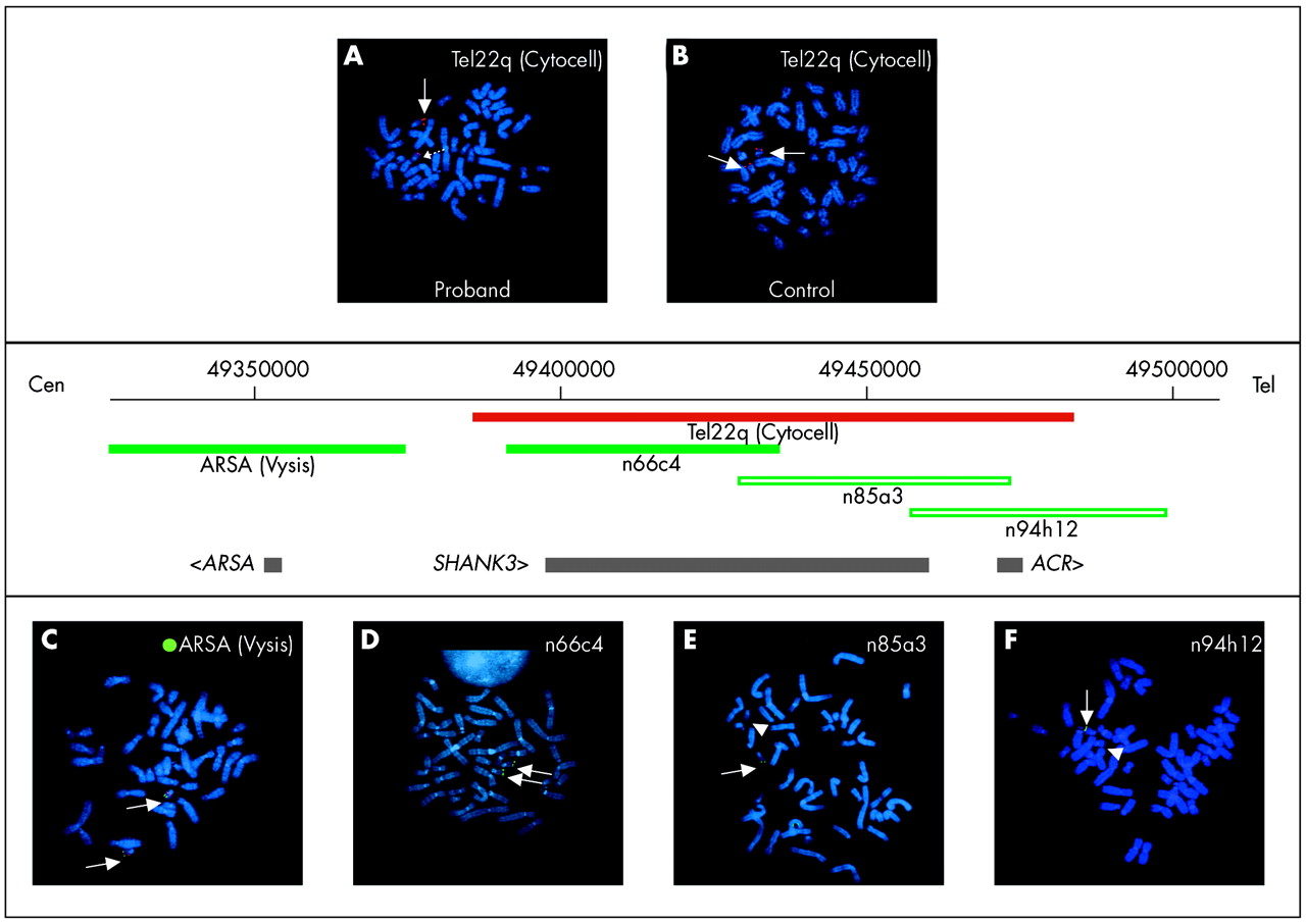
FISH tests with subtelomeric probes (Multi-T, Cytocell) revealed a slight difference in intensity between the signals detected on the two 22q homologous chromosomes, which led us to suspect a partial 22qter deletion. The patient was re-tested with a single Tel22q probe (Cytocell) and FISH analysis confirmed the previous results (fig 1A). FISH with the ARSA probe (Cytocell) showed two equally strong hybridisation signals (fig 1C). To rule out a possible polymorphism, FISH analysis with probe Tel22q was extended to the parents, and a normal hybridisation pattern in all 50 metaphases analysed was observed (fig 1B shows the father’s Tel22q FISH analysis). FISH analysis with cosmid probe n85a3, partially overlapping the Tel22q probe, confirmed the deletion (fig 1E). The deletion breakpoint was refined using the following cosmid clones: n66c4, partially overlapping n85a3, gave a normal hybridisation pattern (fig 1D), while cosmid clones n85a3 (fig 1E), n94h12 (fig 1F), and n1g3 (not shown) were deleted. Thus, the breakpoint was localised in the proximal part of n85a3, which contains the SHANK3 gene, and the size of the deletion was estimated to be 100 kb (fig 1, map). These data overlap those obtained by Anderlid et al.10 FISH analysis on the parents with probes 22q (Cytocell), n66c4, n85a3, 92h12, and n1g3 gave normal results in all 50 metaphases analysed (not shown).

Molecular cytogenetic characterisation of the 22q13.3 deletion. (A) Specific subtelomeric probe for chromosome 22q (Cytocell). Abnormal chromosome 22 (dotted arrow) shows on 22qtel a signal smaller than that detected on the normal homologue (arrow). (B) Normal control FISH for probe Tel22q (Cytocell). The two chromosome 22 homologues (arrow) show equal hybridisation signal intensity. (C) Specific commercial probe for the DiGeorge/VCFS syndrome (Vysis). Proximal signals at 22q11.2 (red) indicate hybridisation of LS1 Tuple 1 and distal signals (green) indicate hybridisation at the LS1 ARSA (arylsulfatase A) locus. Arrows indicate the presence of signals on the distal end of both chromosomes 22. (D) Cosmid probe n66C4. Both chromosome 22 homologues (arrow) show the presence of hybridisation signals. (E, F) Distal cosmid probes n85a3 and n94h12, respectively, show the absence of hybridisation signals on one chromosome 22 (arrowhead). The map of the 22q13.3 deleted region is shown in the centre of the figure: deleted clones (n85a3 and n94h12) are depicted as green empty bars, the retained clones (LS1 ARSA and n66c4) and the Tel22q probe (Cytocell) as green and red bars, respectively. Gene are shown as horizontal grey bars (bottom). The map was constructed according to the UCSC Genome Browser; the localisation of the Cytocell probe is shown according to Anderlid et al.10
To exclude the presence of cryptic imbalances (microdeletion or microduplication) in other locations of the genome, we performed genome-wide array-CGH with an average resolution of 75 kb (Agilent). The array contains oligomers for the ARSA and ACR genes, whereas SHANK3 is not represented. The analysis results were normal apart from the two ACR targets at distal 22q that were deleted (data not shown).
Molecular analysis of our case and that previously reported by Anderlid et al
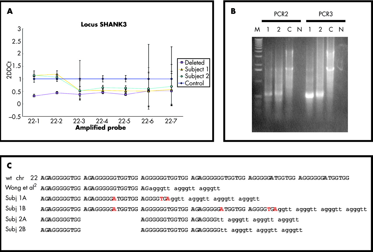
Real-Time PCR quantification showed that a normal control and a deleted control could be easily differentiated at all seven amplicons; both subjects under study appeared to have normal copy number for 22-1 and 22-2, and to be deleted for 22-3 to 22-7 (fig 2A). We used ACP technology14 to clone the deletion breakpoints, selecting chromosome 22 specific primers in the L2 region, immediately adjacent to the D22S163 polymorphism (fig 3).15 Nested amplification generated specific fragments of approximately 500 bp in the two patients only; the patients and a normal control shared a non-specific 1.6 kb fragment (fig 2B). Sequencing of the 500 bp fragments showed truncation of the chromosome 22 sequence and healing by the addition of a telomere repeat array (fig 2C). The two breakpoints are no more than 15 bases apart, within a short simple repeat located between exons 8 and 9 of SHANK3, 900 bp distal to MS607A and approximately 2 kb proximal to MS607B.15 Small variations in the sequence of the terminal nucleotides suggest artifactual recombination between copies of the simple repeat during PCR or telomerase “stuttering” at the start of repeat addition (fig 2C). The overall structure of the SHANK3 gene, together with the position of all known breakpoints, is shown in fig 3.

Breakpoint identification. (A) Real-Time PCR analysis at seven locations (22-1 to 22-7, on the x axis) in the SHANK3 gene. 2DDct values and their standard deviations are shown on the y axis; normal control (Control, blue diamonds), deleted control (Deleted, purple squares), the subject described in Anderlid et al10 (Subject 1, yellow triangles), and the subject described in this paper (Subject 2, light blue circles) are shown. (B) Breakpoint amplification from subject 1 (1), subject 2 (2), a normal control (C), and water control (N) were analysed on a 1% agarose/1× TAE gel; the molecular weight marker (M) is Marker X (Roche Diagnostics, Basel, Switzerland). (C) Comparison between the chromosome 22 breakpoint sequences. Only a portion of the repeat is shown, and single repeat units are divided by spaces. The normal chromosome 22 sequence (wt chr 22) is on the first line; the sequence reported in Wong et al, two types of clones obtained from subject 1 (Subj 1A and 1B), and two types of clones obtained from subject 2 (Subj 2A and 2B) were aligned with the wt sequence. Larger spacing was used for subject 2 in order to optimise the alignment. The telomeric repeats found in all breakpoint sequences are shown in lowercase lettering. Base differences with the wt sequence are typed in red.

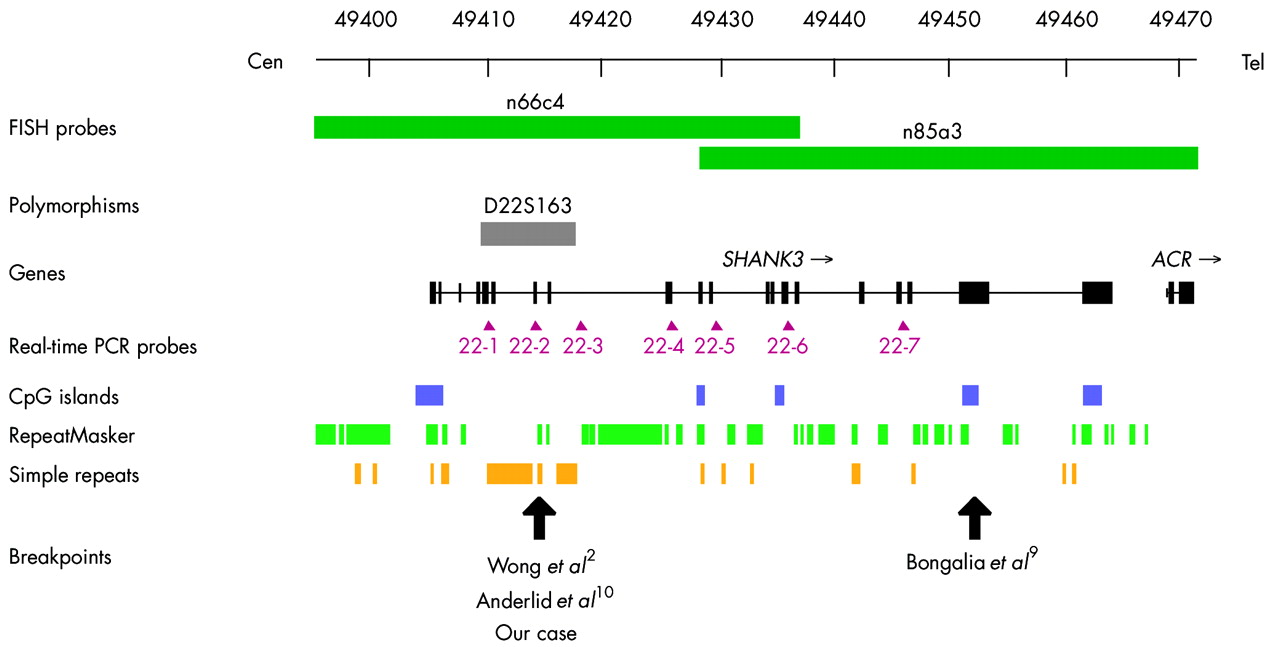
Genomic structure of the human SHANK3 gene. The locations of selected FISH probes, the D22S163 polymorphism, Real-Time PCR probes, selected structural motifs, and all known breakpoints are indicated.
DISCUSSION
Breakpoint analysis
The molecular basis of the mechanisms leading to terminal deletions is poorly defined, essentially for two reasons: first, the breakpoints have been determined at the base pair level in only a few cases; second, the observed breakpoints could be different from the original breakpoints as a consequence of the action of repair mechanisms activated by DNA double strand breakages.16 Molecular analysis of a large cohort of monosomy 1p36 subjects demonstrated that deletion sizes vary widely from about 1 Mb to more than 10.5 Mb in the most distal portion of 1p36 with no single common breakpoint.3 This is also the case with most 22q13 deletions, but in the two cases analysed here the deletion breakpoints fall within SHANK3 no more than 15 bp apart, inside a short simple repeat located between exons 8 and 9 of the gene. The breakpoints of two additional rearrangements involving SHANK3 have been determined so far; in a 22q deletion2 the breakpoint was located within the same 15 bp repeat, while in a t(12;22) translocation9 it was located in exon 21 (see map in fig 3). Our data demonstrate that the region containing the D22S163 polymorphism15 and its flanking repeats constitutes a tight deletion hotspot within the SHANK3 gene. The highly polymorphic locus detected by microsatellite clone cMS607 (D22S163) in MboI digested DNA was described as being composed of two distinct microsatellite arrays, one with moderate heterozygosity (607A) contained in the cMS607 plasmid, and the other (697B), contributing to the greatest part of the variability, not contained in cMS607.15 Our re-analysis based on the published chromosome 22 sequence shows that D22S163 contains only a minisatellite repeat with an 88 bp period (chr 22: 49408021–49411525) and moderate size variability. Most of its heterozygosity is due to variations in the number and location of MboI restriction sites. The shorter simple repeat involved in 22q13.3 recurrent breakpoints lies distally approximately 800 bp from D22S163, while a third, apparently non-polymorphic, minisatellite repeat (chr 22: 49413872–49415514) is located 1.4 kbp downstream of the second repeat. No recombination associated motifs17 were identified in or around the breakpoint region. In addition, Mfold analysis18 did not reveal any structural feature that could explain the hotspot region’s extreme narrowness. An answer may lie in the sequence itself, an imperfect (AGAGGGGGGTGGTGG)10 repeat with a >75% G content. The repeat does not appear to be variable in size, since Southern blot analysis revealed a PvuII band of less than 1 kb in all subjects tested, a size consistent with the 930 bp expected from the chromosome 22 genomic sequence (not shown). However, it is quite refractory to amplification, yielding fragments of variable size and repeat copy number, and sequencing, making it hard to determine with absolute certainty the parental sequence. Thus, although variations in the sequence of the terminal nucleotides (fig 2C) are probably due to recombination between copies of the simple repeat, there is still a chance that other mechanisms, such as errors in sequence repair by telomerase at the start of repeat addition, may be involved.
Telomere healing has been demonstrated in a series of non-recurrent terminal deletions of chromosomes 1p,16 7q,19 16p,20–22 and 22q.19 For the first time, we demonstrate the presence of a hotspot for a recurrent terminal deletion healed by de novo telomere addition.
Genomic rearrangements, such as translocations and gross deletions, have been associated with the presence of non-B DNA conformations at or near the breakpoints.23–25 Some of the sequences involved in the formation of non-B structures are inverted, mirror and direct repeats, left handed Z-DNA, and tetraplex forming sequences.24 The direct repeat involved in the 22q13 deletion is presumably able to form slipped (hairpin) structures, but it also has a strong potential for forming tetraplexes. In fact, using computational methods,26 we can predict that the SHANK3 repeat is about four times more likely to form a tetraplex structure (G-quartet score: 0.666) than the telomeric (T2AG3)n repeat (score: 0.166), where tetraplex formation is well documented.27 In this respect, it is intriguing to speculate that telomerase could be more efficiently recruited to breakpoint sites with a telomere-like structure.
The 22q13.3 syndrome
This deletion syndrome is characterised by neonatal hypotonia, normal to accelerated growth, absent to severely delayed speech, global developmental delay, and minor dysmorphic facial features.6 Additional clinical features suggestive of 22q13.3 deletion include relatively large and fleshy hands, dysplastic toenails, sacral dimple, decreased perspiration,7 and behavioural characteristics consisting of mouthing or chewing non-food items, increased tolerance to pain, and autistic-like behaviour.28 The increasing number of patients being reported supports the hypothesis that this syndrome may be a common source of mental retardation and may be considered the second most common subtelomeric deletion, after the 1p36.6 deletion.3
All three cases with a common breakpoint within SHANK32,10 (this report) share a number of common phenotypic features, such as mental retardation and developmental delay with severely delayed or absent expressive speech. These features have been described in all cases with larger deletions or r(22) chromosomes and in a subject with a t(12;22) balanced translocation involving the SHANK3 gene.9 In addition, characteristic autistic-like behaviour was present in the case described by Anderlid and colleagues and in our case. Neonatal hypotonia was not present in our case or in the cases reported by Anderlid et al10 and Wong et al.2 As already noted,9,13SHANK3 haploinsufficiency is almost certainly responsible for the major neurological and psychiatric features of the 22q13 syndrome, as well as for the regression of skills experienced by many patients.10,13 On the other hand, there are some obvious differences between the cases. In particular, mental retardation was severe in our case but was always mild in the other cases with two small deletions and a translocation. In addition, autistic behaviour was first seen in our subject at the age of 2 years and became more noticeable over time, while the patient described by Anderlid and colleagues10 exhibited abnormal behaviour with autistic features (lack of contact, stereotypic movement) at a later age (late teens). In the case described by Bonaglia et al,9 the VABS test, administered to both parents when the patient was 7 years and 6 months old, demonstrated an overall AE of 39 months. Specifically, the boy showed relative strength in Daily Living Skills and Socialization domains with AEs of 46 and 41 months, respectively, while Motor Skills AE was 37 months and Receptive Language and Expressive Skills AE was 22 months. His behavioural phenotype has some features also found in autistic subjects (stereotypic movements, hyperactivity, hyperkinesia), but the pattern of VABS is not characteristic of autistic disorder.
Thus, subjects with the same kind of SHANK3 disruption can exhibit different degrees of severity in their phenotype.
SHANK3 and neurological deficits
The minimum region of overlap of rearrangements leading to the 22q13 syndrome is a 100 kb region between cosmid n66c4 proximally and cosmid n94h12 distally. The clone n66c4 is distal to the ARSA locus and overlaps the 5′ half of SHANK3, while clones n85a3 and n94h12 overlap the 3′ end of SHANK3 and ACR, respectively (fig 1). SHANK3 belongs to a family of proteins that interact with receptors and structural proteins of the post-synaptic membrane, and is the central link between receptors and the actin cytoskeleton. These proteins are important scaffolding molecules in the post-synaptic density (PSD) and function to receive and integrate synaptic signals and transduce them into the post-synaptic cells. In addition to their role of assembling the PSD during synaptogenesis, they may play a role in synaptic plasticity and in the regulation of dendritic spine morphology.29–31SHANK3 is the best candidate gene for the neurological deficits (developmental delay and absent speech) in the 22q13.3 syndrome since it is located in the critical region, is always deleted in all reported cases with 22q13.3 syndrome, encodes a structural protein located in the PSD, and is involved in spine maintenance in hippocampal neurons.31
Overview of the 22q13.3 deletion syndrome
Approximately 75% of subjects with the 22q13.3 deletion syndrome have pure 22q deletions,13 either terminal or interstitial, and about 25% have deletions resulting from an unbalanced translocation32 or other structural rearrangement13 such as r(22)8 or reciprocal translocations interrupting the SHANK3 gene.9,10
In spite of the increasing number of reported cases, the 22q13.3 deletion remains under-diagnosed due to failure to detect the 22qter deletion in routine chromosome analysis and to recognise the phenotype on clinical examination. As a consequence, its incidence has not yet been established.7
Since the deletions range in size from 100 kb10 to more then 9 Mb,13 high resolution karyotype analysis will miss many of them. FISH analysis with specific subtelomeric probes can also give ambiguous results, and requires careful evaluation of the hybridisation signal intensity; just discriminating between the presence or absence of a signal is not sufficient. The two cases presented here, having a deletion partially overlapping the commercial subtelomeric probe, highlight the difficulties in interpreting the results and indeed suggest that many similar cases may be overlooked. Considering the non-specificity of the phenotype, all subjects presenting global developmental delay and severe speech delay should undergo appropriate tests (FISH, MLPA) in order to search for a cryptic 22q13 deletion. Obviously, the recruitment of additional cases will lead to a better characterisation of the syndrome and to determination of its incidence.
Acknowledgments
The authors wish to thank Giorgia Menozzi and Uberto Pozzoli for their help with computational methods, and Manuela Sironi for critical reading of the manuscript.
REFERENCES
Footnotes
-
Published Online First 11 November 2005
-
↵* The first two authors contributed equally to this work
-
This work was supported by cofin03-MIUR (to OZ), cofin04-MIUR, the FIRB 2001 (to OZ), the Italian Telethon Foundation (GP0247Y01 to OZ), and the Cariplo Foundation (to OZ)
-
Competing interests: none declared