Article Text

Abstract
Sotos syndrome is an overgrowth syndrome characterised by pre- and postnatal overgrowth, macrocephaly, advanced bone age, and typical facial features. Weaver syndrome is a closely related condition characterised by a distinctive craniofacial appearance, advanced carpal maturation, widened distal long bones, and camptodactyly. Haploinsufficiency of the NSD1 gene has recently been reported as the major cause of Sotos syndrome while point mutations accounted for a minority of cases. We looked for NSD1 deletions or mutations in 39 patients with childhood overgrowth. The series included typical Sotos patients (23/39), Sotos-like patients (lacking one major criteria, 10/39), and Weaver patients (6/39). We identified NSD1 deletions (6/33) and intragenic mutations (16/33) in Sotos syndrome patients. We also identified NSD1 intragenic mutations in 3/6 Weaver patients. We conclude therefore that NSD1 mutations account for most cases of Sotos syndrome and a significant number of Weaver syndrome cases in our series.
Interestingly, mental retardation was consistently more severe in patients with NSD1 deletions. Macrocephaly and facial gestalt but not overgrowth and advanced bone age were consistently observed in Sotos syndrome patients. We suggest therefore considering macrocephaly and facial gestalt as mandatory criteria for the diagnosis of Sotos syndrome and overgrowth and advanced bone age as minor criteria.
- NSD1
- overgrowth syndromes
- mutation screening
- genotype/phenotype correlations
Statistics from Altmetric.com
Overgrowth syndromes form a group of heterogeneous conditions resulting from the dysfunction of various processes involving cell proliferation, growth, or apoptosis. Within this group, Sotos syndrome (MIM 117550) is a distinctive condition characterised by the combination of overgrowth and multiple congenital anomalies/developmental delay.1,2 Diagnostic criteria include specific facial features (prominent forehead with receding hairline, downward slanting palpebral fissures, and pointed chin), pre- and postnatal overgrowth, large head circumference, and advanced bone age.3 Variable degrees of mental retardation are usually observed.
Weaver syndrome (MIM 277590) is seen less commonly than Sotos syndrome and is characterised by a typical craniofacial appearance (micrognathia with a deep horizontal chin crease), deep set nails, camptodactyly, and advanced carpal osseous maturation.4 Despite phenotypic differences, several authors have proposed that Sotos and Weaver syndromes could be allelic diseases.5
The two conditions are largely sporadic but autosomal dominant inheritance has been occasionally reported and several chromosomal anomalies have been observed in Sotos patients.6–9 A balanced de novo translocation has been recently reported (t(5;8)(q35;q24.1)),10 and cloning the translocation breakpoint led Kurotaki et al11 to ascribe the disease to large scale NSD1 deletions on chromosome 5 in most of their patients (66%). By contrast, a large majority of point mutations (70%) and only a minority of large deletions (8%) were found in the British population.12 The aim of our study was to systematically screen our series of 33 Sotos and six Weaver syndrome patients for NSD1 deletions and mutations.
METHODS
Patients
A total of 39 patients were included in the study, namely 33 Sotos and six Weaver syndrome patients. In all cases, routine G banding and R banding chromosome analyses showed a normal karyotype with no evidence of deletions or duplications and molecular analyses ruled out fragile X syndrome. All patients were regularly followed (once a year) and had repeated bone age assessment at various ages. Among the 33 Sotos patients, 23 were considered as typical Sotos patients as they fulfilled the diagnostic criteria defined by Cole and Hughes3 (that is, facial gestalt, overgrowth >2 SD, advanced bone age, and macrocephaly >2 SD).3 Ten were considered as Sotos-like patients as they presented with the specific facial gestalt and macrocephaly but were lacking one major criterion, namely advanced bone age and/or overgrowth.13 Finally, six were considered as Weaver syndrome patients. They all presented with the suggestive facial gestalt, overgrowth, macrocephaly, deep set nails, camptodactyly, and accelerated carpal maturation.
Chromosome and FISH analyses
Metaphase spreads were prepared from phytohaemagglutinin stimulated peripheral blood lymphocyte cultures using standard procedures of hypotonic treatment and methanol/acetic acid fixation (3:1). RHG and GTG banding analyses were performed according to standard protocols.14 FISH probes were labelled with biotin-16-dUTP or digoxigenin-11-dUTP (Boehringer-Mannheim) using a commercially available random priming kit (Gibco-BRL). Biotin labelled probes were detected using Texas Red (TR) conjugated to avidin and digoxigenin labelled probes were detected using fluorescein isothiocyanate (FITC) conjugated to anti-digoxigenin. Slides were counterstained with 4′, 6′-diamidino-2-phenylindole (DAPI). Image capture and analyses were performed using a Zeiss Axiophot epifluorescence microscope equipped with the appropriate filter combination for detecting TR, FITC, and DAPI. The images were captured by a cooled CCD camera using an image analysis system (Vysis). Ten hybridised metaphases were analysed for each probe.
Search for NSD1 deletions
Blood samples from probands and their parents were obtained and genomic DNA was isolated from EDTA anticoagulated blood by a salting out procedure. To screen for NSD1 deletions, we searched for the unbalanced inheritance of the chromosomal region encompassing the NSD1 gene. Three polymorphic microsatellite markers (namely 5q35/CA1, 5q35/CA2, and 5q35/CA3) were identified within the sequence of PAC clones CTC-340P19 (GenBank accession number AC027317), CTC-286C20 (GenBank accession number AC027314), and RP11-265K23 respectively (GenBank accession number AC110005). Marker 5q35/CA1 is located 200 kb upstream of the NSD1 coding region while markers 5q35/CA2 and 5q35/CA3 are intragenic (fig 1). Fluorescent genotyping was performed as previously described.15

Physical map spanning the NSD1 gene. Arrows indicate genes and rectangles correspond to PAC clones. Positions of the microsatellite markers are indicated.
Sequence analyses of the NSD1 gene
Based on the predicted genomic sequence, 31 primer pairs were used for PCR amplification of exons and splicing junctions of the NSD1 gene (table 1). PCR products were purified with Exo-SAP (Amersham) and directly sequenced on an ABI PRISM 3100 DNA Sequencer (Perkin Elmer-Applied Biosystems) using the Dye Terminator method according to the manufacturer’s instructions.
Primers and PCR conditions for NSD1 analysis
RESULTS
NSD1 deletions
Two intragenic NSD1 microsatellite markers and one marker located 200 kb upstream of the NSD1 gene (but within the commonly deleted region) were tested in the probands and their parents. In 6/33 Sotos patients, hemizygosity for at least one marker was found (table 2), while all other patients were heterozygous for at least one marker. None of the parents was found to carry a NSD1 deletion, suggesting that all deletions occurred de novo. All deletions were largely similar in size and were of paternal origin. In all cases, the deletion was subsequently confirmed by FISH analysis. None of the Weaver syndrome patients had a NSD1 deletion.
Genotype analyses at the NSD1 locus in six Sotos syndrome cases
Mutation analyses of the NSD1 gene
Sotos/Weaver syndrome patients with balanced biparental contribution at the NSD1 locus were further analysed for intragenic NSD1 mutations by direct sequencing. A total of 16 different mutations were found in 27/33 Sotos patients. The mutations spanned the whole coding sequence and, in most cases, were likely to result in a truncated inactive NSD1 protein. Indeed, mutant genotypes included frameshift insertions or deletions (8/16), nonsense (4/16) and missense mutations (4/16, table 3). Interestingly, 2/4 missense mutations were located within the SET domain, a highly conserved functional domain of the protein. We also identified distinct deleterious NSD1 mutations in 3/6 Weaver patients, including a frameshift mutation, a nonsense and a missense mutation affecting the SET domain. All mutations were found to have occurred de novo, as they were not found in the parents of affected subjects. The mutations were not identified in 50 controls.
NSD1 mutations identified in Sotos and Weaver patients
Finally, we identified nine polymorphisms including four conservative and four non-conservative changes. The non-conservative changes were found in both patients carrying pathogenic NSD1 mutations and in healthy parents.
Genotype/phenotype correlations
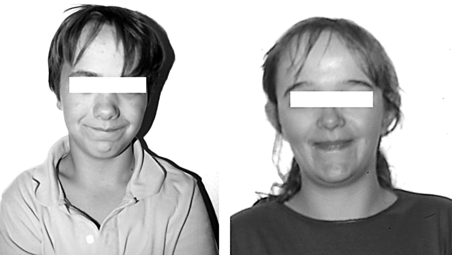
Table 4 summarises the clinical features in the six Sotos patients carrying NSD1 deletion and in the 16 patients with point mutations. All patients had a typical facial gestalt (fig 2A,B) with macrocephaly and ventriculomegaly, regardless of the mutant genotype. In addition, a significant advance of the paternal age at conception was observed.
Clinical manifestations in the Sotos syndrome children carrying a mutation at the NSD1 locus

Facial features in Sotos patients with NSD1 deletion (top) or mutation (bottom). Note the prominent forehead, downward slanting palpebral fissures, long face, and pointed chin.

Facial features in Sotos patients with NSDI mutation.
Among the six Sotos patients carrying a NSD1 deletion, 2/6 were typical Sotos patients and 4/6 were considered as Sotos-like patients based on an overgrowth of 1.5-2 SD and the absence of advanced bone age. Moreover, 4/6 were severely mentally retarded with no speech at all, behavioural problems and no ambulation at 4 years of age. Additional features included congenital heart defects (3/6, septal defects, persistent ductus arteriosus or Ebstein malformation), feeding difficulties (3/6), macrosomia, neonatal hypoglycaemia, seizures, and corpus callosum agenesis (2/6). A true cutis laxa was present in one child with a renal malformation. A cleft palate was present in one patient and a labial cleft in another one.
Among the 16 Sotos patients carrying NSD1 point mutations, 14/16 were typical Sotos patients and 2/16 were considered as Sotos-like patients because of the absence of advanced bone age. Variable degrees of mental retardation with a marked delay in the acquisition of verbal skills were observed. Additional features included hyperlaxity (9/16), macrosomia (8/16), deep set nails (7/16), strabismus and scoliosis (6/16), febrile convulsions and feeding difficulties (3/16), neonatal hypoglycaemia, jaundice, and septal defect (2/16). Table 5 and fig 3 show the clinical features and the facial appearance of the Weaver patients carrying a NSD1 point mutation.
Clinical manifestations in the Weaver syndrome patients carrying a NSD1 anomaly

Facial features in two Weaver patients with a NSD1 mutation. Note the broad forehead and deep horizontal chin crease.
DISCUSSION
Studying a series of 33 Sotos children, we found evidence of mutant NSD1 genotypes in more than 66% of our patients. Mutant genotypes included deletions (18.2%) and point mutations (48.5%). Interestingly, NSD1 deletions were consistently of paternal origin with a significant advance of the paternal age at conception in our series. All point mutations described here were hitherto unreported de novo mutations, and most of them were truncating mutations. Taken together, our results support the view that NSD1 anomalies are a major cause of Sotos syndrome. They are in agreement with the British study but at variance with the Japanese study, where an extremely high rate of NSD1 deletions was observed.9,10 The reason for these discrepancies remains to be elucidated. One possible hypothesis is an increased susceptibility to deletions related to putative repetition polymorphisms within the Japanese population.
From a clinical viewpoint, it is worth noting that macrocephaly and facial gestalt were consistent features in all patients carrying a mutant NSD1 genotype. By contrast, overgrowth and advanced bone age were not consistently observed. The advanced bone age is probably not a consistent finding in Sotos syndrome. We suggest therefore considering macrocephaly and facial gestalt as mandatory criteria for the diagnosis of Sotos syndrome while overgrowth and advanced bone age should be regarded as minor criteria.
Comparing the clinical phenotype of children carrying either a deletion or a mutation, we failed to detect distinctive features except for the severity of mental retardation. Indeed, 4/6 children carrying a NSD1 deletion were extremely severely mentally retarded with no language at all, major delay in motor milestones, and autistic features. By contrast, in patients carrying NSD1 mutations, mental retardation was usually mild to moderate with verbal skills being more affected.
On the other hand, all three Weaver syndrome children carrying NSD1 mutations presented with typical features of the syndrome, and no clinical difference from those Weaver children without NSD1 anomalies could be found. Similar observations have been made by Douglas et al.12 Also, why NSD1 mutations (missense, frameshift, or nonsense mutations) caused either Sotos or Weaver syndromes remains unexplained. Finally, we were unable to find NSD1 anomalies in 50% of Weaver syndrome children or in 33% of Sotos syndrome patients. These features could result from either locus heterogeneity or our current inability to detect all mutations at the NSD1 locus.
We conclude that the NDS1 locus at present accounts for most cases of Sotos syndrome (66.6%) and Weaver syndrome (50%). Ongoing studies will hopefully decide whether these conditions are indeed genetically homogeneous or if another locus is involved in these overgrowth syndromes.