Article Text

Abstract
MWS is a multiple congenital anomaly syndrome, first clinically delineated by Mowat et al in 1998. Over 45 cases have now been reported. All patients have typical dysmorphic features in association with severe intellectual disability, and nearly all have microcephaly and seizures. Congenital anomalies, including Hirschsprung disease (HSCR), congenital heart disease, hypospadias, genitourinary anomalies, agenesis of the corpus callosum, and short stature are common. The syndrome is the result of heterozygous deletions or truncating mutations of the ZFHX1B (SIP1) gene on chromosome 2q22.
- Mowat-Wilson syndrome
- Hirschsprung disease
Statistics from Altmetric.com
In 1998, Mowat et al1 described six patients with a mental retardation syndrome recognised by its characteristic facial appearance in association with Hirschsprung disease (HSCR). One of their patients had a cytogenetic deletion of 2q22–23 and they noted a previously published patient with a 2q22 deletion and similar clinical features. Based on this they proposed that this syndrome was either caused by microdeletion in chromosome 2q22–2q23 or a de novo mutation of a gene within this region. In 2001, the cause of this syndrome was found to be deletions or intragenic mutations of the ZFHX1B gene.2,3 To date, 45 microdeletion/mutation positive cases have been reported.2–10 Several other clinical reports published before the ZFHX1B gene discovery describe patients with syndromic HSCR who probably also have this syndrome.11,12 All mutation positive cases show a similar facial appearance to the original patients.1–12 Although most patients were ascertained on the basis of HSCR, several series have now reported mutations in patients without HSCR.4,6,8,10 Recognition of the characteristic facies with or without Hirschsprung disease (HSCR) has important implications for genetic counselling. All reported cases of this syndrome have been sporadic, resulting from de novo deletion or heterozygous mutation of the ZFHX1B gene. It is important to distinguish this syndrome from the Goldberg-Shprintzen syndrome,16 which has some clinical overlap but may have an autosomal recessive basis. Although the molecular basis of Goldberg-Shprintzen syndrome has not yet been established, characterisation of the ZFHX1B gene suggests clinical and genetic heterogeneity for the phenotypes of HSCR associated with mental retardation and microcephaly.
CLINICAL FEATURES (TABLE 1)
Clinical features in patients with deletion or mutation of the ZFHX1B gene
Facial phenotype
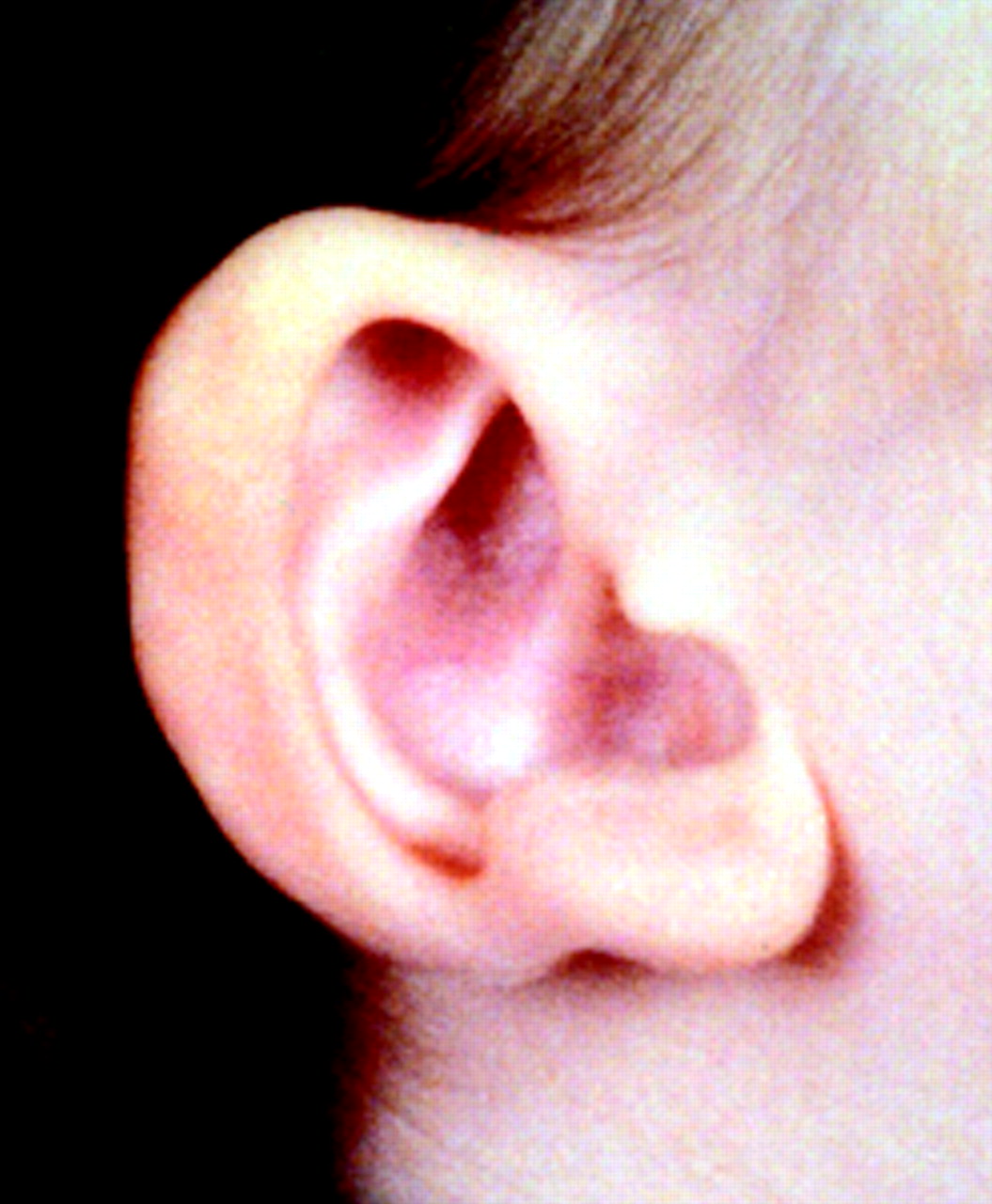
All our cases and those published cases where a photograph is provided show similar facial features. In infancy (fig 1), there is a square shaped face with a prominent but narrow triangular chin, hypertelorism, deep set but large eyes, broad nasal bridge, saddle nose, prominent, rounded nasal tip, open mouth, full or everted lower lip, posteriorly rotated ears, and large uplifted ear lobes with a central depression. The configuration of the ear lobes, which have been described as like “orechiette pasta” or “red blood corpuscles” in shape, is a consistent and easily recognisable feature (fig 2).


Characteristic appearance of the posteriorly rotated ears with an uplifted ear lobe and central depression (“corpuscular-like”).
In childhood (fig 3A), the face lengthens and the chin becomes more prominent. The eyebrows are often broad and horizontal with a wide medial separation. The columella becomes more obvious. The vermilion border often has an upper case “M” shape, such that the upper lip is full centrally but thins rapidly laterally. The children commonly have a smiling, open mouthed expression, and drooling is a significant feature in some. In children with blue eyes, patchy clumps of darker iris pigment occur, prompting the description of heterochromia of the irides.

Facial appearance of case 158 (425 C to S142X) in childhood (A) and adolescence (B).
In adolescents and adults the face is long with prognathism and a long, pointed or “chiselled” chin. The nasal tip lengthens and overhangs the philtrum, while the upper-mid portion of the nasal profile becomes convex (fig 3B).
Neurological
All subjects with MWS have at least moderate and usually severe intellectual disability, although formal IQ studies have not generally been reported. In our experience, most patients have a happy demeanour with frequent smiling. Speech is absent or restricted to a few words and is disproportionately delayed compared to comprehension. Some patients communicate successfully with signing. The children are usually hypotonic in the first few years of life, with delayed motor milestones. Typically they will cruise or stand holding on to furniture but are late to walk independently. The mean age of walking in our series was ~4 years but a proportion remained non-ambulatory. The gait is wide based and the arms are often held flexed at the elbows, with hands up. This stance, combined with the smiling face, has led to a suggested diagnosis of Angelman syndrome in some patients.
Most patients have had seizures (90%) or an abnormal EEG. The onset of seizures is usually in the second year of life, although they may begin in infancy or late childhood. The seizures can be varied in nature although myoclonic seizures have not been seen. In some cases the seizures have been resistant to treatment in childhood, but appear to be more easily managed in adolescents and adults.
Growth/musculoskeletal
Growth parameters at birth, including head circumference, are usually in the normal range. Postnatal short stature (< or =3rd centile) is usual, although several patients have normal stature, and one of our adult patient’s height is on the 90th centile. Microcephaly is sometimes present at birth, but more often becomes apparent in infancy. Not all patients are microcephalic.
Most subjects are of slender build. The fingers are slender and tapered in childhood, with prominence of the interphalangeal joints developing in adolescence and adulthood. The feet usually show mild calcaneovalgus deformity and long toes. One patient has broad halluces and another has unilateral duplication of the hallux.8
Hirschsprung disease
Hirschsprung disease was present in 28 of the 45 published cases (table 2). The penetrance for HSCR in males was 65% (11/31), for females 57% (8/14), and 62% for the total group. The male:female ratio of those with HSCR was 2.5:1, and in those without HSCR was 1.8:1. The data on the length of the aganglionic segment is incomplete in published reports. Long segment and short segment HSCR is described in males and females. Females may be more likely to have long segment HSCR than males.8 The male preponderance of published cases may be because of biased ascertainment, as males may be more likely to manifest HSCR.13 Of the 17 cases without HSCR, five are reported to have severe constipation not investigated by rectal biopsy. It is possible that these patients could have very short segment HSCR, as was the case in one of our patients.1,3 It is now well established that ZFHX1B mutations can lead to a mental retardation/MCA syndrome without HSCR,4–6,8,10 but it is likely that at present the syndrome is under-recognised in this group.
Sex ratio and penetrance for HSCR in MWS patients
Congenital heart disease
Congenital heart disease has been reported in 45% of patients with MWS. The heart anomalies reported include patent ductus arteriosus (5), atrial septal defect,(4) ventricular septal defect (3), tetralogy of Fallot (3), pulmonary atresia (1), pulmonary stenosis (3), aortic coarctation (2), bicuspid aortic valve (1), and aortic valve stenosis (1).1,4–6,8,9,15
Genitourinary anomalies
Genitourinary anomalies are common in MWS. Anomalies described include hypospadias (9), “webbed penis” (3), bifid scrotum (2), cryptorchidism/undescended testes (6), duplex kidney (1), pelvic kidney (1), vesicoureteric reflux (5), and hydronephrosis (2). Hypospadias is a common finding in males. In 24 patients where this information was available, hypospadias was present in 60% of patients (15/25). So far, no girls with renal tract anomalies have been reported, but many have not had the relevant imaging studies.1,4,5,7,8,15
Cerebral structural anomalies
Microcephaly is a common but not invariable feature, present in 84% of reported cases. Cerebral anomalies described include total or partial agenesis of the corpus callosum (13/31=42%), cerebral atrophy (7), poor hippocampal formation, and frontotemporal hypoplasia (3) with temporal dysplasia. Not all published cases have had cranial imaging so these findings may be under-represented.1,4–6,8,15
Other clinical features
Bifid uvula/submucous cleft was reported in three of our patients. A high arched palate is often present, possibly secondary to hypotonia. Pyloric stenosis has been reported in three patients. Early “nystagmus” owing to fixation difficulties is frequently reported in infancy, but this resolves. Many patients have convergent strabismus. One patient has unilateral ptosis but none has an ocular coloboma. Some patients with blue irides have shown dark pigment clumps in the irides, described as heterochromia irides by some authors. No patient has had sensorineural deafness. Wilson et al8 described one patient who gradually developed widespread “raindrop” depigmentation in the truncal region, of uncertain cause.
DIFFERENTIAL DIAGNOSIS
The series reported by Amiel et al5 suggests that there is genetic heterogeneity for syndromic HSCR as only eight of the 19 patients they selected with microcephaly/MR/HSCR were found to have deletion or mutation of the ZFHX1B gene. Goldberg-Shprintzen syndrome (GSS) is another form of syndromic HSCR with microcephaly and MR.16 Although there is considerable clinical overlap, the spectrum of the clinical features and the facial dysmorphism in GSS appear to be different from MWS. Features more commonly described in GSS include cleft palate, ptosis, synophrys, arched eyebrows, curled eyelashes, and ocular coloboma.11,12,14,16–22 The underlying basis of GSS may be autosomal recessive, as sib recurrence and consanguinity is reported, but the molecular basis remains unclear. So far, no information about ZFHX1B gene analysis in patients with definite GSS has been reported.
Other conditions causing mental retardation and HSCR include chromosomal deletions involving loci of known HSCR causing genes (del 10q11, del 13q22) and Waardenburg type IV (SOX10), Smith-Lemli-Opitz, Bardet-Biedl, and BRESHEK syndromes.13 Patients with HSCR and hypospadias should have 7-dehydrocholesterol levels performed to exclude Smith-Lemli-Opitz syndrome.
Some patients with MWS have been thought to have Angelman syndrome,23 based on the presence of severe MR, seizures, ataxia, microcephaly, prominent jaw, and a happy behavioural phenotype. One patient with unilateral duplication of the hallux and agenesis of the corpus callosum was previously diagnosed as having the acrocallosal syndrome.8
THE ZFHX1B GENE
Mowat-Wilson syndrome was initially localised to the chromosome 2q22–23 region, based on two patients with interstitial deletions in this region.1,9 In 2001, Wakamatsu et al2 identified the SIP1 gene (later designated ZFHX1B) within a 5 kb microdeletion associated with a translocation involving 2q22 in a patient with characteristic features and confirmed its causative role by identification of intragenic mutations in three further patients with similar syndromic HSCR. Cacheux et al3 independently identified a patient with a (2;11) translocation disrupting the SIP1 gene, and found intragenic mutations in a further four cases, including three of the originally reported patients.1,15 Amiel et al5 screened 19 patients with HSCR/MR/microcephaly, selected from cohort of 250 patients with HSCR, and found microdeletions or mutations of the ZFHX1B gene in eight. All of the patients with identified mutations had the typical dysmorphic features and seven had HSCR. Yamada et al6 described mutations in six patients with the dysmorphic phenotype, but without HSCR. Zweier et al4 identified mutations in two patients without HSCR and Wilson et al8 described a further six patients without HSCR. These reports confirmed that HSCR is not an essential feature of mutations in ZFHX1B. Finally, several patients have now been reported with the typical clinical syndrome, with and without HSCR, where no abnormality involving ZFHX1B can be shown.6,8
ZFHX1B, located on chromosome 2q22, spans approximately 70 kb of DNA, consists of 10 exons and 9 introns, and encodes a protein product, Smad interacting protein 1 (SIP1), consisting of 1214 amino acids. SIP1 is a member of the δEF1/Zfh-1 family of two handed zinc finger/homeodomain transcription factors involved in the TGF-β/BMP/Smad mediated signalling cascade.24 SIP1 shows 97% homology to the mouse form (Sip1) at the amino acid level, with exactly the same functional domains.3 SIP1 contains a Smad binding domain (SBD), a homeobox-like domain (HD), interspersed with a zinc finger cluster, and two other separated zinc finger clusters (ZF) at the amino and carboxy terminals.25ZFHX1B mRNA is detected in nearly all human tissues (heart, brain, placenta, lung, liver, skeletal muscle) as well as mouse heart and brain.3 SIP1 expression is also detected in fetal human tissues including brain, kidney, liver, and spleen. Mouse Sip1 is expressed in many brain regions (hypothalamus, cerebellum, cortex, rhombencephale, hippocampus, mid brain, and spinal cord). As a transcription modulator, SIP1 is likely to have a crucial role in embryonic development.26–28 XSIP1, the Xenopus homologue of SIP1, has been shown to be active in early embryogenesis. It is strongly expressed early in prospective neuroectoderm, then in the neural plate, and later in the neural tube and neural crest.3,30,31 SIP1 interacts with other Smad proteins 2, 3, and 5.30 Activated Smad proteins form hetero-oligomeric complexes that translocate to the nucleus to control the expression of target genes in a cell specific manner. SIP1 appears to be a promoter of invasion in malignant epithelial tumours. It acts by downregulating E-cadherin transcription via binding to both conserved E2 boxes of the minimal E-cadherin promoter.32 The mechanism underlying the phenotypic effects of SIP1 mutation has not yet been determined. Understanding this process may provide insights into the development of the enteric nervous system and the cause of intellectual disability. Interestingly Espinosa-Parrilla et al33 recently showed the lack of SMADIP1 expression in the developing human heart even though many patients have congenital heart defects.
GENOTYPE-PHENOTYPE CORRELATIONS
MWS has been associated with heterozygous deletions, translocations, and intragenic mutations of ZFHX1B (table 3). All but one of the intragenic mutations are nonsense or frameshift mutations, expected to produce a truncated or absent protein, suggesting that haploinsufficiency leads to an altered gene dosage effect at critical stages of early development (fig 4). One patient has a splice site mutation (splice site G to A +1 exon 2) which causes complete loss of normal splicing. This causes a total absence of splicing, producing a protein which has retained the intron (exon 2-intron 2-exon 3). This changes the reading frame leading to a stop at the next nonsense codon, resulting in a truncated protein. The mutations are located throughout the coding region of the gene; although 58% of mutations are in exon 8, this comprises around 60% of the coding sequence.
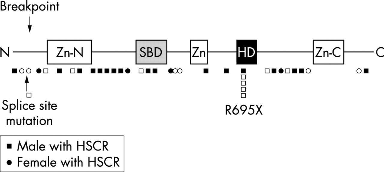
Schematic representation of the SIP1 protein showing the predicted sites of protein truncation from identified mutations. Zn=N amino terminal zinc finger, SBD=Smad binding domain, Zn=zinc finger, HD=homeobox domain, Zn-C=carboxy terminal zinc finger.
All seven patients (three males and four females), with deletion of the entire ZFHX1B gene have HSCR,1,5,6,9 as does the patient with a translocation disrupting the gene in intron 2. Only one of the five patients with R695X (the only recurrent mutations so far reported) has HSCR. Of the 32 other intragenic mutations, 16 are in patients with HSCR, but there is no clear genotype-phenotype correlation in respect to location of the mutation and penetrance of the HSCR phenotype. Zweier et al4 showed persistence of truncated SIP1 mRNA in a patient with a (nt553–554 ins TG) in exon 5, indicating that mRNA decay and complete haploinsufficiency of SIP1 may not be present in all cases. No other studies of this nature have been reported. There are no patients with missense mutations to help determine the effects of disruption of specific domains within SIP1. There are no data available to suggest that some of the deleted patients have a similar sized deletion. The presence of genomic repeats flanking the ZFHX1B gene to account for the deleted patients has not been determined. It is not known whether the parental origin of the deleted or mutated ZFHX1B gene has an effect on phenotype as these data have not been provided.
MWS without ZFHX1B mutation
Several series have now reported typical patients (diagnosed by clinicians familiar with the phenotype), who do not have demonstrable mutations in ZFHX1B. These include one patient of Yamada et al6, three patients of Amiel et al,5 and two further patients from our cohort (fig 5).5,6,8,15 These patients may have undetected abnormalities involving ZFHX1B, but genetic heterogeneity for this syndrome cannot be completely excluded, in particular mutations affecting alternative genes acting on the same developmental pathways.

Example of a female without detectable mutation as an infant (A) and in early adolescence (B).
GENETIC COUNSELLING
All cases of MWS have been sporadic, caused by de novo deletions or new dominant mutations in ZFHX1B. Where the diagnosis is certain, in a sporadic patient, families can be counselled that the recurrence risk is low, although the possibility of germline mosaicism cannot be excluded.