Article Text

Abstract
Background: Biallelic germline mutations in the mismatch repair genes MLH1, MSH2, MSH6 or PMS2 cause a recessive childhood cancer syndrome characterised by early-onset malignancies and signs reminiscent of neurofibromatosis type 1 (NF1). Alluding to the underlying genetic defect, we refer to this syndrome as constitutional mismatch repair-deficiency (CMMR-D) syndrome. The tumour spectrum of CMMR-D syndrome includes haematological neoplasias, brain tumours and Lynch syndrome-associated tumours. Other tumours, such as neuroblastoma, Wilm tumour, ovarian neuroectodermal tumour or infantile myofibromatosis, have so far been found only in individual cases.
Results: We analysed two consanguineous families that had members with suspected CMMR-D syndrome who developed rhabdomyosarcoma among other neoplasias. In the first family, we identified a pathogenic PMS2 mutation for which the affected patient was homozygous. In family 2, immunohistochemistry analysis showed isolated loss of PMS2 expression in all tumours in the affected patients, including rhabdomyosarcoma itself and the surrounding normal tissue. Together with the family history and microsatellite instability observed in one tumour this strongly suggests an underlying PMS2 alteration in family 2 also.
Conclusion: Together, these two new cases show that rhabdomyosarcoma and possibly other embryonic tumours, such as neuroblastoma and Wilm tumour, belong to the tumour spectrum of CMMR-D syndrome. Given the clinical overlap of CMMR-D syndrome with NF1, we suggest careful examination of the family history in patients with embryonic tumours and signs of NF1 as well as analysis of the tumours for loss of one of the mismatch repair genes and microsatellite instability. Subsequent mutation analysis will lead to a definitive diagnosis of the underlying disorder.
Statistics from Altmetric.com
The highly conserved mismatch repair (MMR) system in organisms corrects replication errors in newly synthesised DNA. In humans, mismatches and small insertion-deletion loops (IDLs) are detected by one of two heterodimers, MSH2•MSH6 (MutSα) or MSH2•MSH3 (MutSβ). MutSα is involved in the repair of base/base mismatches and misalignments of one or two nucleotides, whereas MutSβ recognises larger IDLs. MutSα (or MutSβ) recruits a second heterodimer, MLH1•PMS2 (also named MutLα), which possesses an endonuclease active site and allows MutLα to introduce random nicks at sites spanning the mismatch. Subsequent loading of EXO1 at the 5′ side of the mismatch leads to activation of its 5→3′ exonuclease activity, leading to the removal of the error-containing DNA fragment. The repair process is finalised by polymerase δ and its cofactors proliferation cell nuclear antigen (PCNA) and replication factor C (RFC), which fill in the single-stranded gap. In a final step, ligase I seals the remaining nick (reviewed by Jiricny1). In addition to DNA repair activity, the MMR system is also involved in apoptotic response to a variety of DNA-damaging agents (reviewed by Jiricny2), and human PMS2 deficiency is associated with impaired immunoglobulin class-switching recombination.3
Heterozygous germline loss-of-function mutations of the genes encoding the crucial components of this MMR system (MLH1, MSH2, MSH6 or PMS2) cause Lynch syndrome, a well characterised dominant cancer syndrome associated with hereditary non-polyposis colorectal cancer (HNPCC) and other malignancies (reviewed by Peltomaki4). Tumours arising in these patients result from somatic loss of the remaining wild type MLH1, MSH2, MSH6 or PMS2 allele, which leads to impaired MMR and accumulation of somatic mutations.
To date, some 46 families with patients harbour biallelic germline mutations of MLH1, MSH2, MSH6 or PMS2 have been reported (reviewed by Wimmer and Etzler5). Constitutive biallelic inactivation of one of these mismatch repair genes causes a recessive childhood cancer syndrome characterised by early-onset malignancies, café-au-lait spots (CLS) and/or other signs of neurofibromatosis type 1 (NF1). Alluding to the underlying genetic defect, we refer to this syndrome as constitutional mismatch-repair-deficiency (CMMR-D) syndrome.
The tumour spectrum of the reported patients with CMMR-D syndrome includes primarily haematological neoplasias, brain tumours and Lynch syndrome-associated tumours.5 To date, other tumours, such as neuroblastoma, Wilm tumour, ovarian neuroectodermal tumour or infantile myofibromatosis, have been found only in individual cases. Herein, we report on two families with CMMR-D syndrome. In both families one affected individual developed rhabdomyosarcoma (RMS), suggesting that RMS is part of the CMMR-D syndrome tumour spectrum.
CASE REPORTS
Family 1
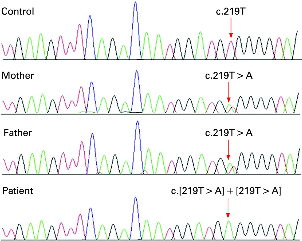
The index patient was a 9-year old boy born to consanguineous parents. He was diagnosed with embryonal RMS in the left nasolabial fold at the age of 3 years and was treated according to the recommendations of the German Soft Tissue Sarcoma Study Trial CWS96. He relapsed 1 year later and was treated according to the high-risk arm of the CWS-2002-P protocol. At the age of 8 years, he required emergency surgery due to intussusception of the colon. The transverse colon was resected and a primary end-to-end anastomosis was performed. Histological investigations showed the diagnosis of a single adenocarcinoma. Lymph nodes were negative and he received FOLFOX (folinic acid, fluorouracil, oxiplatin) treatment. After the fourth cycle, the patient developed rectal bleeding and was seen at our institution for a second opinion. Notably, physical examination found several café-au-lait spots ,and rectal endoscopy showed no polyps or tumours. A similar case with multiple CLS and several childhood cancers due to a biallelic mutation of PMS2 was recently diagnosed at our institution6 and, thus, we suspected the same syndrome in this patient. Immunohistochemistry or microsatellite instability analysis of the tumours was not possible because the patient underwent surgery before presenting at our institution and a tumour specimen was unavailable. Therefore, informed consent for germline mutation analysis was obtained from the parents, and PMS2 analysis was performed using published RNA-based methods.7 Sequencing of reverse transcriptase PCR products revealed a novel homozygous mutation, c.[219T→A]+[219T→A], leading to a premature stop codon (p.Cys73X) in PMS2 exon 3. This finding was confirmed by sequencing of genomic DNA from the patient and is consistent with the diagnosis of CMMR-D syndrome due to a homozygous PMS2 mutation. As expected, both parents were heterozygous carriers of the mutant PMS2 allele (fig 1). Mutation analysis of DNA from two healthy siblings and members of the extended family was not possible. The absence of malignancies in the heterozygous parents is not surprising as heterozygous PMS2 mutations are known to have a low penetrance.8 9 The mother’s father died from a bone tumour at the age of 60 years and her brother had lung cancer. Two of the father’s cousins were diagnosed with brain tumours at the ages of 21 and 50 years, respectively, and another cousin was diagnosed with bladder carcinoma at the age of 55 years.

Sequencing results in family 1 showing heterozygosity for the mutation c.219T→A (p.C73X) in the parents and homozygosity in the patient.
Family 2
A 22-year-old woman presented with a 3-year history of anemia and a weight loss of 9 kg over the previous few months. A large abdominal mass was identified on ultrasonography and CT scans. Colonoscopy identified four synchronous adenocarcinomas of the rectum, sigmoid, transverse colon and caecum. The patient underwent total proctocolectomy with en bloc Whipple resection. Histopathological examination confirmed the four primary colorectal cancers and identified six additional tubulovillous and villous adenomas in the rectum. The patient was also noted to have multiple CLS.
Because of her family history and cutaneous features the patient was referred to the Familial Gastrointestinal Cancer Registry (FGICR) for genetic evaluation. Immunohistochemistry of the MLH1, MSH2, MSH6 and PMS2 proteins showed complete lack of PMS2 expression in both normal and tumour tissue, but normal expression of the other proteins. Microsatellite instability analysis of BAT25 and BAT26 in two representative tumours showed instability at BAT25 in one tumour and stability in the other. BAT26 was stable in both tumours. Collectively, these data suggested underlying biallelic PMS2 germline mutations in the patient. Informed consent for germline mutation analysis was obtained by the patient. Unfortunately, it was not possible to amplify long-range PCR products in genomic DNA extracted from blood lymphocytes10 and RNA extracted from puromycin-treated lymphocytes of the patient was not available for cDNA sequencing.7 Hence, no reliable germline PMS2 analysis was possible.
The proband’s sister was initially diagnosed with a left occipital anaplastic astrocytoma at the age of 16 years. Four months after the astrocytoma diagnosis, she was found to have an undifferentiated sarcoma of the right pterygoid fossa. Molecular analysis of the tumour did not reveal features consistent with alveolar rhabdomyosarcoma, Ewing sarcoma, or desmoplastic small round cell tumour, therefore the pathology suggested some embryonal RMS-like features. Initially, she was referred to a genetics clinic to be evaluated for Li–Fraumeni syndrome. The family declined genetic testing for TP53 and refused DNA banking. The patient died at the age of 18 years of her brain tumour. Retrospectively performed immunohistochemistry showed complete lack of PMS2 expression in the astrocytoma and the sarcoma (fig 2) as well as normal tissue of the patient, confirming further the suspected biallelic PMS2 germline mutations in both sisters.
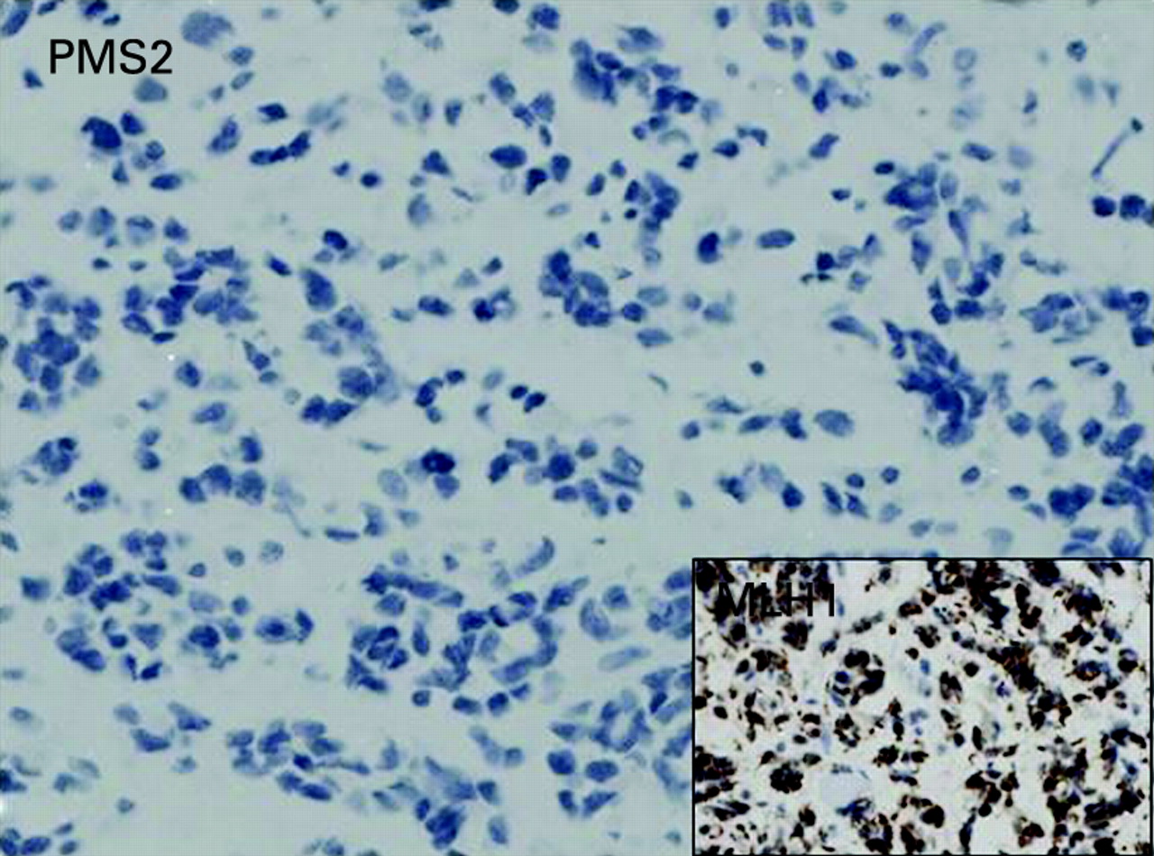
Immunohistochemistry analysis showed isolated lack of PMS2 expression in the rhabdomyosarcoma from patient 2 in family 2. The mismatch repair protein MLH1 (shown in the inset) as well as MSH2 and MSH6 stained positive in the same tumour.
The sisters’ parents are second cousins, and an extended family history showed complex consanguinity (fig 3). The mother of the children reportedly died of colorectal cancer at 28 years of age, a maternal uncle died of leukaemia at 9 years and another maternal uncle died of a brain tumour at 19 years. Because of the complex consanguinity, there may have been biallelic PMS2 mutation carriers in the mother’s generation also. Confirmation of the cancers was not possible as the family members were treated in other countries.

Pedigree of family 2. The index patient is marked with an arrow. The sites of tumours and the age at diagnosis in years (y) are indicated below the symbols. The sister of the index patient developed rhabdomyosarcoma at the age of 16 years.
DISCUSSION
Together these patients show that RMS is part of the tumour spectrum of CMMR-D syndrome. Notably, RMS has been described previously in a patient who was closely related to three people in whom a homozygous MLH1 mutation was detected,11 12 suggesting that this individual also has CMMR-D. Hence, the spectrum of malignancies occurring in patients with CMMR-D syndrome may be extended from haematological neoplasias, brain tumours and LS-associated tumours to embryonic tumours, such as embryonic RMS and possibly also Wilm tumour13 14 and neuroblastoma.15
RMS is the most common soft tissue sarcoma in children. Cancer syndromes predisposing to RMS include Li–Fraumeni syndrome, Costello syndrome, familial retinoblastoma and Beckwith-Wiedemann syndrome.16 An increased incidence of RMS has also been reported for NF1.17 As in the two reported cases, the histological subtype tends to be embryonal RMS in patients with NF1.17 18 We speculate that, because of the clinical overlap between CMMR-D syndrome and NF1, some people with RMS and a clinical (mis)diagnosis of NF1 actually have CMMR-D syndrome. The cancer and family history of at least one of these patients strongly suggest this possibility.5 19 Homozygosity of all tested microsatellite markers in the NF1 locus may indicate parental consanguinity in another reported NF1 case with RMS, rendering CMMR-D syndrome a possible alternative diagnosis also in this patient.20 Identifying the underlying genetic alteration in patients with CMMR-D syndrome is important, as it has implications also for the wider family. There is a recurrence risk of 25% for the recessive disorder in the family and heterozygous carriers have an increased risk for Lynch syndrome-related tumours. Careful examination of the family history of the patient, analysis of the tumour(s) for loss of one of the mismatch repair proteins and microsatellite instability, and subsequent mutation analysis will allow a definitive diagnosis of the underlying disorder in patients with RMS and signs of NF1.
REFERENCES
Footnotes
Competing interests: None.