Article Text

Abstract
In the last two decades the extraordinary advances in molecular biology of arrhythmogenic right ventricular cardiomyopathy/dysplasia (ARVC/D) have provided significant insights into our understanding of the disease aetiology by showing that it is a genetic disorder of the cardiac desmosomes and that interactions between mechanical disruption of cell–cell adhesion and defects of desmosomal-mediated intracellular signalling are likely to be involved in the pathogenesis of the ARVC/D phenotype. The discovery of the causative genes for ARVC/D offers the possibility of identifying genetically-affected individuals before potentially malignant clinical phenotype occurs. Moreover, the evaluation of abnormal localisation of desmosomal proteins by immunohistochemical analysis on endomyocardial biopsy samples represents a promising test for ARVC/D diagnosis. Early detection of ARVC/D and preventive therapy of young individuals at highest risk of experiencing sudden cardiac death may be improved by molecular genetic screening within affected families and may alter the clinical management of patients. At present, however, the clinical use of genotyping is limited by the incomplete knowledge of causative mutations and the complex genetic background of the disease, which accounts for the incomplete penetrance and the marked variability of the phenotype expression. This review addresses the advances in the molecular biology of ARVC/D, with particular reference to the genetic basis of the disease, and how these advances have impacted on understanding the disease pathogenesis, on diagnosis and in establishing management strategies.
- Arrhythmic right ventricular dyplasia
- implantable cardioverter defibrillator (ICD)
- sudden cardiac death
- ventricular fibrillation
- genetics
Statistics from Altmetric.com
- Arrhythmic right ventricular dyplasia
- implantable cardioverter defibrillator (ICD)
- sudden cardiac death
- ventricular fibrillation
- genetics
Introduction
Arrhythmogenic right ventricular cardiomyopathy/dysplasia (ARVC/D) is an inherited disease of the heart muscle that predominantly affects the right ventricle (RV). The main pathological feature is the progressive loss of RV myocardium and its replacement by fibrofatty tissue.1–5 The condition was initially believed to be a developmental defect of the RV myocardium, leading to the original designation of ‘dysplasia’.6 This concept has evolved over the last 25 years into the current perspective of a genetically-determined ‘cardiomyopathy’.7 8 Clinical manifestations develop most often between the second and fourth decade of life and are related to ventricular tachycardia (VT) or ventricular fibrillation (VF) which may lead to sudden death, primarily in young people. Progression of RV muscle disease and left ventricular (LV) involvement may result in right or biventricular heart failure.2 3 Ventricular arrhythmias are worse during or immediately after exercise, and participation in competitive athletics has been associated with an increased risk of sudden death.9 10 Clinical diagnosis of ARVC/D is often difficult due to the non-specific nature of the disease features and the broad spectrum of phenotypic manifestations, ranging from severe to concealed forms.11–13
Advances in the molecular genetics of ARVC/D have provided new insights into the understanding of the pathogenesis and pathophysiology of the disease, showing that in its pure form it is a genetic disorder resulting from defective desmosomal proteins.14 15 The availability of molecular testing for screening known gene mutations offers the possibility to identify genetically-affected individuals by DNA characterisation.14–17 The potential clinical impact of genotype determination includes early diagnosis with prediction of clinical phenotype, arrhythmic risk stratification and therapeutic interventions aimed at preventing sudden death. Although available, genetic testing for ARVC/D is not yet routinely used in the clinical setting for a number of reasons.
The prevalence of causal genes and mutations has yet to be determined, so a negative genetic test does not exclude the possibility that the phenotype is due to a mutation of unknown and thus untested disease-causing genes.18 The certainty of detecting causative mutation carriers (and non-carriers) is further limited by the difficulty in distinguishing causative mutations (mostly missense gene variants) from polymorphisms as well as by the potential presence of an undetected second pathogenetic mutation in the same or another gene. Most importantly, there is no evidence that the genotype may help with management strategies. Molecular genotyping is currently applied to relatives of a proband with a known mutation probably associated with ARVC/D for risk assessment (presymptomatic test), while it is not routinely used in isolated cases with a borderline phenotype for confirming the diagnosis (diagnostic test).
Abnormal expression of genetically defective desmosomal proteins at intercalated discs has recently been proposed as a potential biomarker for ARVC/D diagnosis by immunohistochemical analysis of endomyocardial biopsy samples.19 20 However, further research is needed to validate its diagnostic accuracy before such a test can be considered for clinical use.
This review provides an up-to-date perspective on the molecular biology of ARVC/D, with particular reference to the advances made in defining the genetic basis of the disease and how these advances have impacted on understanding disease pathogenesis, on diagnosis and in establishing management strategies.
Advances in molecular biology
Aetiology
The estimated prevalence of ARVC/D in the general population ranges from 1 in 2000 to 1 in 5000.15–17 A familial background has been demonstrated in 30–50% of cases of ARVC/D but, in the remaining ‘isolated’ cases, an underlying familial disease with incomplete penetrance or limited phenotypic expression cannot be excluded. ARVC/D affects men more frequently than women, with a ratio ranging from 1.3 to 2.4.21 22
The inherited nature of ARVC/D has been recognised since 1982 when Marcus et al16 described 24 adult cases, two in the same family. In a 1988 report on eight Italian families, the autosomal dominant pattern of inheritance with incomplete penetrance and variable expression was demonstrated for the first time.23
The first chromosomal locus (14q23-q24) for autosomal dominant ARVC/D was published in 1994 after clinical evaluation of a large Venetian family.24 Subsequently, linkage analysis provided evidence for genetic heterogeneity with sequential discovery of several ARVC/D loci on chromosomes 1, 2, 3, 6, 10, 12, 14 and 18 (table 1). An autosomal recessive variant (so-called Naxos disease), in which there is a cosegregation of cardiac (ARVC/D), skin (palmoplantar keratosis) and hair (woolly hair) abnormalities, has been mapped on chromosome 17 (locus 17q21).25 The discovery that ARVC/D is a cell–cell junction disorder came from a molecular genetic study of this ‘cardiocutaneous’ condition and stimulated the research for other related genes. Epidermal cells and myocytes share similar mechanical junctional apparatus (desmosome and fascia adherens) and are exposed to high frictional stress. This suggested that common genes encoding proteins of intercellular junctions were proper candidates for a molecular genetic study aimed at identifying disease-causing mutations. Intercellular junctions are composed of a core region, which mediates cell–cell adhesion, and a plaque region which provides attachment to the intermediate filament cytoskeleton. There are three major groups of desmosomal proteins: (1) transmembrane proteins (ie, desmosomal cadherins) including desmocollins (DSC) and desmogleins (DSG); (2) desmoplakin (DSP), a plakin family protein that binds directly to intermediate filaments (desmin in the heart); and (3) linker proteins (ie, armadillo family proteins) including plakoglobin (JUP) and plakophilins (PKP) which mediate interactions between the desmosomal cadherin tails and DSP.26 A schematic representation of the desmosomal complex is shown in figure 1.
Chromosomal loci and disease-causing genes in arrhythmogenic right ventricular cardiomyopathy/dysplasia (ARVC/D)

Schematic representation of the intracellular and intercellular components of the desmosomal plaque. Three separate families of proteins assemble to form desmosome: desmosomal cadherins (desmoglein and desmocollin), armadillo proteins (plakoglobin and plakophilin) and plakins (desmoplakin). The desmosomal cadherins present with extracellular domains that play a pivotal role in cell adhesion whereas the intracellular domain interacts with the armadillo proteins. Among the latter, plakophilin binds to the N-terminal domain of desmoplakin and the C-terminal of desmoplakin anchors desmin intermediate filaments. IF, intermediate filaments; PM, cytoplasmic membrane. Modified from Basso et al.5
The first ARVC/D-causing gene, the JUP gene, was identified by McKoy et al14 in patients with Naxos disease. The gene encodes desmosomal protein JUP, which is the major constituent of the cell adhesion junction. A recessive mutation of DSP has been found to cause another cardiocutaneous syndrome, so-called Carvajal disease, characterised by biventricular involvement.27 Mutations in desmosomal protein genes have also been shown to cause the more common (non-syndromic) autosomal dominant form of the disease. DSP was the first defective gene to be associated with autosomal dominant ARVC/D by Rampazzo et al.28 Subsequently, a variety of mutations in plakophilin-2 (PKP-2) have been identified in almost one-third of unrelated probands from three different cohorts of patients with ARVC/D across the world.29–31 In familial cases of ARVC/D, mutations in two additional desmosomal genes have been reported—desmoglein-2 (DSG-2) and desmocollin-2 (DSC-2) genes.32 33 This consistent type of protein alteration supports the concept of a ‘final common pathway’ of genetically-determined cardiomyopathies, ARVC/D being deemed to be a desmosomal disease, hypertrophic cardiomyopathy a sarcomeric disease and dilated cardiomyopathy a cytoskeletal disease, with some exceptions.8 34
Autosomal dominant ARVC/D has been linked to other genes unrelated to the cell adhesion complex. Mutations in the gene encoding for cardiac ryanodine receptor (RyR2), which is responsible for calcium release from the sarcoplasmic reticulum, have been identified in four families with an autosomal dominant form of ARVC/D characterised by effort-induced polymorphic VT associated with segmental RV cardiomyopathic changes.35 A defective transforming growth factor β3 (TGFβ3) gene has been reported in all clinically affected members of a large ARVC/D family.36 Mutations resulting in overexpression of the TGFβ3 gene are expected to induce a myocardial fibrosis response by stimulating mesenchymal cells to proliferate and to produce extracellular matrix components. Moreover, it has been shown that TGFβ3 modulates expression of genes encoding desmosomal proteins in different cell types, so defective genes may affect cell–cell junction stability as well. The most recently discovered ARVC/D gene is TMEM43 which causes a highly lethal and fully penetrant disease variant (ARVD5).37 Little is known about the function of the TMEM43 gene, which may be part of an adipogenic pathway regulated by PPARg whose dysregulation may explain the progressive fibrofatty replacement of the myocardium in ARVC/D.
Pathogenesis
According to the widely accepted ‘defective desmosome’ hypothesis, genetically-determined disruption of desmosomal integrity is the key factor leading to the development of ARVC/D.14 20 29 38 39 Although desmosomes are traditionally considered specialised structures which provide mechanical attachment between cells, they are emerging as mediators of intracellular and intercellular signal transduction pathways.40 Ultrastructural investigation in gene-positive ARVC/D patients revealed intercalated disc remodelling with desmosomal abnormalities (ie, decreased desmosomal number, increased desmosomal length and pale desmosomes) and intercellular gap widening, which are strongly in keeping with a cell–cell junction cardiomyopathy.38 39 The mechanism whereby mutations affecting components of the cardiac desmosome result in ARVC/D remains to be elucidated. It is believed that the lack of the protein or the incorporation of defective proteins into cardiac desmosomes may provoke detachment of myocytes at the intercalated discs, particularly under mechanical stress conditions.14 20 29 As a consequence, there is progressive myocyte degeneration and death with subsequent repair by fibrofatty replacement. Rather than being a continuous process, disease progression might occur during periodic bursts (‘hot phases’) in an otherwise stable disease.5 13 15 These disease exacerbations can be clinically silent in most patients but sometimes can be characterised by the appearance of chest pain and life-threatening arrhythmias. Environmental factors such as physical exercise or inflammation might facilitate disease progression by worsening cell adhesion disruption.
Life-threatening ventricular arrhythmias may also occur either during the ‘hot phase’ of myocyte death as abrupt VF or later in the form of scar-related macro-reentrant VT.41
Impaired mechanical coupling might account for abnormal electrical coupling through gap junction remodelling.42 Immunohistochemical and electron microscopy studies on myocardium from patients with Naxos disease revealed reduced localisation of mutant JUP to cell–cell junctions, diminished expression of the gap junction protein connexin-43 and decreased number and size of gap junctions.43 Likewise, in Carvajal syndrome, the immunoreactive signal for both DSP and JUP was markedly diminished at intercalated discs, as was the signal for desmin and connexin-43.44 More recently, similar changes in the various intercalated disc proteins were observed in the classic form of ARVC/D without cardiocutaneous manifestations due to PKP-2 mutations.45 46 These preliminary findings suggest that gap junction remodelling might provide an additional mechanism for conduction delay and RV electrical instability which may result in potentially fatal arrhythmias before fibrofatty myocardial replacement occurs histologically.
The ‘defective desmosome’ hypothesis was also supported by transgenic animal models which in part reproduced the ARVC/D phenotype. A transgenic mouse with cardiac restricted overexpression of the C-terminal mutant (R2834H) DSP has been shown to develop increased cardiomyocyte apoptosis, myocardial fibrosis and lipid accumulation as well as biventricular dilation/dysfunction.38 Another study on heterozygous JUP-deficient mice showed that mutant animals had increased RV volume, reduced RV function and more frequent and severe VT of RV origin.47 In this animal model, endurance training accelerated the development of RV dysfunction and arrhythmias. However, the clinical phenotype of this heterozygous JUP-deficient mutant mouse showed only limited similarity to the human forms of ARVD/C because none of the mutant mice had myocardial fibrofatty replacement and only inconsistent RV dilation was noted.
Data from Garcia-Gras et al40 on DSP-deficient mice suggest an alternative molecular mechanism of disease which implicates inhibition of the canonical Wnt/β-catenin signalling through Tcf/Lef transcription factors in the pathogenesis of ARVC/D. In this study, cardiac-specific loss of the desmosomal protein DSP was sufficient to cause nuclear translocation of JUP, increased expression of adipogenic and fibrogenic genes and the development of a ARVC/D-like phenotype consisting of myocardial fibrofatty infiltration, cavity enlargement and ventricular arrhythmias. This evidence for potential Wnt/β-catenin signalling defects implicates a role for cell adhesion proteins, not only as passive players in providing mechanical attachment between cells but also as regulators in cardiac development, in myocyte differentiation and in the maintenance of the myocardial architecture.
Further insights into the pathophysiological mechanisms involved in ARVC/D (those initiating myocardial damage) are provided by a study of transgenic mice (Tg-NS) with cardiac overexpression of DSG-2 gene mutation N271S-dsg2.48 Transgenic mice reproduced the clinical features of ARVC/D, including spontaneous ventricular arrhythmias, cardiac dysfunction, biventricular dilation with aneurysms and sudden death at young age. Investigation of transgenic lines with different levels of transgene expression attested to a dose-dependent dominant-negative effect of the mutation. The study showed for the first time that myocyte necrosis is the key initiator of myocardial injury. Myocyte necrosis was the first manifestation of disease in all Tg-NS hearts studied. Electron microscopic evaluation in 2–3-week-old Tg-NS/H mice showed disruption of the sarcolemma, disgregation of myofilaments and other cytoplasmic elements and mitochondrial swelling, all ultrastructural features consistent with cardiomyocyte necrosis. Myocardial cell death subsequently triggers an inflammatory response and massive calcification within the myocardium, followed later by injury repair with fibrous tissue replacement and myocardial atrophy.
Subcellular redistribution of desmosomal proteins
Saffitz and coworkers used immunohistochemistry to characterise the expression, location and redistribution of mutant and non-mutant desmosomal proteins in patients with ARVC/D. By first studying patients with recessive cardiocutaneous syndromes such as Naxos disease and Carvajal syndromes, they showed that the mutant JUP protein, although clearly expressed in the patient tissue, failed to localise normally at intercellular junctions as also did its desmosomal binding partners.19 20 This suggests that a mutation in a single desmosomal protein may alter the subcellular distribution from desmosomes to intracellular pools of other proteins which are assembled to form intercellular junctions. An immunohistochemical study of patients with dominant ARVC/D showed that gene mutations lead to abnormal localisation of desmosomal proteins even when the mutation affects only a single allele. This finding indicates that the genetically-defective protein may alter binding interactions of one or more desmosomal proteins with redistribution from junctional to cytosol and, potentially, with changes in nuclear signalling and gene expression patterns. These observations have provided an opportunity for developing a new diagnostic test for ARVC/D based on immunohistochemical analysis of human myocardial samples aimed to identify changes in the distribution of desmosomal proteins (figure 2).19
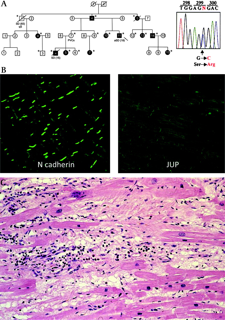
Immunoreactive plakoglobin (JUP) signal and histological features in a sudden death victim from familial arrhythmogenic right ventricular cardiomyopathy/dysplasia (ARVC/D) caused by a mutant desmoplakin (DSP) gene. (A) Family pedigree of the ARVC/D family and identified mutation (S299R) in exon 7 of the DSP gene. (B) Immunohistochemical analysis of human myocardial samples of the proband who died suddenly at the age of 15 years showing a marked reduction in immunoreactive signal levels for JUP but normal signal levels for the non-desmosomal adhesion molecule N-cadherin. (C) Histology of the ventricular myocardium showing ongoing myocardial atrophy with early fibrofatty replacement.56
Contributing role of viruses
At histological examination the typical ARVC/D features of fibrofatty myocardial replacement changes, particularly during the acute ‘bursts’ of disease progression, are frequently associated with inflammatory infiltrates which probably play a role in triggering life-threatening arrhythmias.2 3 Whether the inflammatory cells are a reaction to cell death or the consequence of infective or immune mechanisms is not known. Cardiotropic viruses such as adenovirus, hepatitis C virus and parvovirus B19 have been reported in the myocardium of some patients with ARVC/D, and they have been proposed as possible aetiological agents, thus supporting an infective pathogenesis.49 However, the viral agent might be just an innocent bystander or play a secondary but still important role.50 According to the latter hypothesis, the genetically dystrophic myocardium could favour viral settlement (superimposed myocarditis) leading to progression or the precipitation of the disease phenotype. It is noteworthy that similar pathological features of inflammation have been described in spontaneous animal models of ARVC/D, with a clinical picture dominated by right heart failure and ventricular arrhythmias with the risk of sudden death.51 Moreover, the recent demonstration of massive inflammatory cell infiltrates following acute myocyte necrosis in the early stages of the disease onset in a DSG-2 transgenic animal model supports the reactive nature of myocarditis in ARVC/D.48
Clinical impact of molecular biology
The discovery of gene mutations involved in the pathogenesis of ARVC/D has offered the potential to increase the accuracy of diagnosing ARVC/D by genotyping. The potential clinical utility of gene mutation screening in ARVC/D is primarily based on the ability to confirm the diagnosis in a proband with a clinical suspicion of ARVC/D, to distinguish true ARVC/D from phenocopies, to identify genetically-affected family members at a preclinical stage and to reassure unaffected individuals, and to stratify the risk of sudden death.15–17 The available studies indicate that molecular genetic diagnosis by screening for mutations in desmosomal genes is feasible in clinical care and successful in a proportion of patients.
Diagnostic genetic test
Diagnostic genetic testing is applied to probands with a clinical suspicion of ARVC/D with the aim of establishing a conclusive diagnosis. Identification of desmosomal gene mutations may also be useful in the differential diagnosis between ARVC/D and phenocopies such as idiopathic RV outflow tract VT, dilated cardiomyopathy with biventricular involvement, inflammatory cardiomyopathy and cardiac involvement in skeletal muscular dystrophies or sarcoidosis.13 17
Clinical diagnosis
The phenotypic diagnosis of ARVC/D is difficult because of several problems with the specificity of the ECG abnormalities, different potential aetiologies of ventricular arrhythmias with a left bundle branch block morphology, assessment of the RV structure and function and interpretation of endomyocardial biopsy findings. In 1994 an international Task Force proposed criteria to facilitate and standardise the clinical diagnosis of ARVC/D (table 2).11 The proposed strategy consists of reaching a clinical diagnosis by combining multiple sources of diagnostic information such as genetic, ECG, arrhythmic, morphofunctional and histopathological findings. Since their publication, the Task Force criteria have been extremely useful in providing a common approach to the clinical diagnosis of ARVC/D. The criteria were initially designed to guarantee an adequate specificity for ARVC/D among index cases with overt clinical manifestations. The Task Force guidelines have actually helped cardiologists to avoid misdiagnosis of ARVC/D in patients with dilated cardiomyopathy or idiopathic RV outflow tract VT. However, the diagnostic criteria have been shown to lack sensitivity for identification of early/minor phenotypes, particularly in the setting of familial ARVC/D. Accordingly, a revision of the guidelines has recently been undertaken with the goal of improving diagnostic sensitivity but with the important requisite of maintaining diagnostic specificity.12 A comparison of the original versus the revised Task Force diagnostic criteria is shown in table 2. The approach of classifying structural, histopathological, ECG, arrhythmic and genetic features of the disease as major and minor criteria has been maintained. According to the revised criteria, a definite diagnosis of ARVC/D is fulfilled by two major or one major and two minor criteria or four minor criteria from different categories; a borderline diagnosis by one major and one minor or three minor criteria from different categories; and a possible diagnosis by one major or two minor criteria from different categories.12
Original and revised task force criteria for ARVC/D diagnosis
As far as ECG and arrhythmic features are concerned, in the revised Task Force criteria T wave inversion in V1–V3 as well as VT with a left bundle branch block morphology with superior/indeterminate QRS axis, either sustained or non-sustained, have become major criteria. The following findings have been included among the minor criteria: (1) T wave inversion in V1 and V2 in the absence of right bundle branch block (and from V1 to V4 in the presence of complete right bundle branch block); (2) positivity of any one of the three signal-averaged ECG parameters for late potentials; (3) premature ventricular beats >500 per 24 h on Holter monitoring. Furthermore, a limitation of the original Task Force criteria was the lack of quantitative cut-off values for proper grading of RV dilation/dysfunction and fibrofatty myocardial replacement.12 The revised guidelines for diagnosing ARVC/D provide quantitative imaging and histopathological measurement cut-off points for defining normal RV and categorising the various degrees of morphofunctional RV abnormalities.12 Finally, in the revised Task Force diagnostic guidelines the family history criteria have been modified to include molecular genetic information. The identification of a pathogenetic gene mutation in a first-degree relative has become a major criterion for a diagnosis of ARVC/D.
In the original Task Force criteria LV involvement was deemed to be the result of disease progression which occurred late and concomitantly to widespread RV disease, leading to congestive heart failure.7 13 Subsequently, both clinical and autopsy studies showed that LV involvement is a common finding in ARVC/D and may occur earlier than previously thought.2 3 52 53 Gadolinium-enhanced magnetic resonance has been shown to be the gold standard test for identifying LV involvement, even early/minor.5 13 54 55 LV late enhancement predominantly involves the inferolateral and inferoseptal regions and, unlike subendocardial distribution of ischaemic scar, is characteristically localised in the subepicardial or mid-wall layers, similar to the histological pattern of RV fibrofatty myocardial replacement found at post-mortem examination.2 3 Prominent LV late enhancement with ventricular dilation/dysfunction and no or mild RV involvement may be observed in the ‘left dominant’ form of ARVC/D.53 Other clinical markers of this disease variant include ECG abnormalities suggesting LV involvement such as T wave inversion in the leads exploring the LV (V5,V6, L1 and aVL) and ventricular arrhythmias of LV origin (with a right bundle branch block morphology). Genotyping of familial ‘left dominant’ ARVC/D demonstrated the same genetic defect underlying classic ARVC/D—that is, desmosomal gene mutations with a particular role for the gene encoding for DSP.52 56 57 The frequent and early finding of LV involvement in ARVC/D and the identification of a predominantly left-sided variant of the disease led to the current concept that ARVC/D is a genetically-determined heart muscle disease affecting both ventricles (‘arrhythmogenic cardiomyopathy’).
Genetic diagnosis
At present the role of molecular genotyping for diagnosing ARVC/D of index cases with ‘borderline’ or ‘possible’ ARVC/D is limited by the complex genetic background of the disease which accounts for the low penetrance and variable expression of the ARVC/D phenotype and explains our current difficulty in identifying true disease-causing gene defects.15–18 The PKP-2 gene has been most frequently associated with the disease, with a reported prevalence of mutations ranging from 10% to 47% among unrelated probands and reaching 70% among familial ARVC/D cases.17 However, in an Italian series of 80 unrelated ARVC/D probands undergoing genetic screening of all known ARVD/C genes, 13 (16%) carried a DSP mutation, 11 (14%) a PKP-2 mutation, 8 (10%) a DSG-2 mutation and 2 (2.5%) a TGFβ3 mutation.32 Pooled results from major molecular genetic studies of desmosomal genes allow estimation of the overall rate of successful genotyping to approximately 40%.17 The most commonly defective ARVC/D gene is PKP-2 (10–45%), followed by DSP (10–15%), DSG-2 (7–10%) and DSC-2 (2%). Of note, screening for non-desmosomal genes marginally affects the mutation detection rate.17 32 Family studies appear to indicate that different gene defects may be associated in part with peculiar clinical features. For instance, DSP gene mutations generally account for earlier and more severe LV involvement.52 53 56
Most recent genotype–phenotype correlation studies suggest that missense mutations of desmosomal genes, mostly the PKP-2 gene, although relatively common in ARVC/D probands, are not pathogenetic per se and a second gene mutation is often necessary for the disease phenotype to be manifest.58 59 In the study by Xu et al,59 16 of 38 ARVC/D probands (42%) carrying PKP-2 gene mutations showed concomitant variants of the same gene (compound heterozygosity) or of a second desmosomal gene (digenic heterozygosity). The observation that single ‘nonsense’ or ‘frame shift’ mutations of PKP-2 often did not result in clinical disease in the studied families indicates that haploinsufficiency for this gene is not critical or sufficient to cause ARVC/D. Finally, harbouring more than one gene mutation affected not only the disease penetrance but also the degree of phenotypic expression (ie, earlier onset or more severe disease manifestations). The genetic heterogeneity of ARVC/D and the compound/digenic heterozygosity of causative mutations complicate the mutation screening test since all five desmosomal genes must be sequenced, increasing both overall cost and complexity of analysis.
However, approximately half of ARVC/D probands do not harbour any known causative gene mutations. Hence, although a positive test result can be informative, a negative test does not exclude the possibility that the ARVC/D phenotype is due to a mutation in a gene that was not tested. Moreover, the possibility of the presence of a second mutation in the same or another pathogenetic gene (still unknown) curbs the ability to identify conclusively and reassure non-mutation carriers. Moreover, there are desmosomal gene mutations for which too little evidence exists to classify them as disease-causing. In this regard, a number of published PKP-2 and DSG-2 gene mutations, which were originally believed to be pathogenetic, were subsequently identified in 0.5–13.9% of healthy controls and currently classified as polymorphisms.59–61 Although the identification of such gene mutations is of uncertain clinical relevance, the current perspective is that, although not sufficient to cause ARVC/D, they may influence the risk of disease development in positive carriers. According to these genotyping limitations, a positive result for a molecular genetic test of index patients may support a clinical diagnosis but is not conclusively diagnostic while a negative test is non-contributory.15–17
Molecular autopsy
Clinical screening of the surviving relatives of victims of unexplained sudden death may identify ARVC/D in 7–9% of cases.62 63 In the index victim, ARVC/D is usually not diagnosed because of incomplete protocol of morphological investigation or non-qualified autopsy examination. On the other hand, arrhythmic cardiac arrest might theoretically occur in patients with very ‘early stage’ disease before fibrofatty myocardial substitution becomes evident on histological examination. In these cases a diagnosis of ARVC/D necessarily depends on molecular genetic testing (‘molecular autopsy’) as in victims of sudden death from cardiac ion channel diseases. The role of molecular autopsy in ARVC/D is, however, restricted by the above limitations for the confirmatory diagnosis in living index cases.
Presymptomatic genetic diagnosis
The major advantage of genetic screening is the possibility of making a preclinical diagnosis of ARVC/D in the setting of a familial disease. Gene mutation analysis and cascade screening of relatives offers an alternative strategy to serial non-invasive cardiovascular evaluation of families. It is important to remember that molecular genetic testing may only support a clinical overt or suspicious diagnosis but cannot make a diagnosis of ARVC/D itself.16 17 In fact, mutation carriers may either have no disease phenotype (incomplete penetrance) or present clinically with various degrees of clinical manifestations (variable expression) ranging from asymptomatic family members with concealed RV structural abnormalities and no arrhythmias to patients experiencing cardiac arrest or undergoing cardiac transplantation because of RV or biventricular heart failure. Inheriting a mutation does not mean that the individual will present with clinical manifestations of the disease. This suggests that a positive genetic result can only be part of a more comprehensive clinical approach combining multiple sources of diagnostic information such as ECG changes, ventricular arrhythmias and RV morphofunctional/histopathological changes as well as clinical and molecular genetic findings.11 12
The most important clinical impact of genotyping families with ARVC/D is the possibility of identifying genetically-affected relatives who do not yet have signs or symptoms of the disease but have inherited the risk of developing the ARVC/D phenotype on the basis of family history/known causal mutation. It is noteworthy that clinical manifestations of ARVC/D usually develop during adolescence and young adulthood and are preceded by a long preclinical phase. Cardiac arrest often occurs in previously asymptomatic young adults and competitive athletes as the first manifestation of the disease.1–5 16 64 Young age is the most powerful independent predictor of VF in patients with ARVC/D. All efforts should be made to genotype and manage younger family members with ARVC/D, who carry the highest risk of VF, before a malignant phenotype occurs. Early diagnosis of ARVC/D by molecular genetic analysis is warranted because it allows focused surveillance and a specific prevention strategy to be established based on lifestyle modifications such as refraining from participation in competitive sport, closer clinical follow-up aimed at timely identification of alarming symptoms, ECG/echocardiographic abnormalities or ventricular arrhythmias and appropriate prophylactic therapy with β-blockers, amiodarone and/or an implantable cardioverter-defibrillator (ICD) to prevent sudden death.16
Immunohistochemical diagnosis
Preliminary clinical data on the immunohistochemical analysis of cardiac tissue samples show that a reduced signal for JUP at intercalated discs typically occurs in patients with ARVC/D and appears to be an accurate disease biomarker. Asimaki et al19 compared endomyocardial biopsy samples from patients with ARVC/D and myocardium obtained at autopsy from controls; all ARVC/D samples (but no control samples) showed a marked and diffuse (both in the RV and LV) reduction of immunoreactive signal levels for JUP at the intercalated disc, while signal levels for the non-desmosomal adhesion molecule N-cadherin were normal. Other desmosomal proteins showed variable changes. Moreover, in the myocardium obtained from subjects with hypertrophic, dilated or ischaemic cardiomyopathies, the levels of N-cadherin and JUP signals at junctions were indistinguishable from those in control samples. Compared with clinical Task Force criteria, the immunohistochemical test appeared to be highly sensitive (91%) and specific (82%) for diagnosing ARVC.19 However, more recent data from Saffitz and coworkers indicate that a reduced desmosomal protein signal is not specific for ARVC/D as it is also found in patients with sarcoidosis or giant cell myocarditis.65 Thus, the test accuracy for diagnosing ARVC/D may be lower than originally reported and deserves further clinical validation.
Prognosis
Although ICD confers optimal protection against sudden death in patients with ARVC/D, the significant rate of inappropriate interventions and complications—as well as the psychological repercussions (mostly in the younger age group)—argue strongly against indiscriminate implantation of devices. However, risk stratification of affected patients is still incomplete. The available data, based on autopsy series or retrospective clinical studies, suggest that young age, prior cardiac arrest, fast and poorly tolerated VT with different morphologies, syncope, severe RV dysfunction and LV involvement with heart failure are potential predictors of sudden death and worse outcome.4–6 66 Malignant family history seems not to be an important predictor of increased risk in ARVC/D. Figure 3 shows the arrhythmic risk stratification pyramid and the current indications for ICD implantation based on the available data from observational studies on ICD therapy.41 67 68 69

Arrhythmic risk stratification pyramid and current indications for implantation of an implantable cardioverter defibrillator (ICD) based on observational studies on ICD therapy in arrhythmogenic right ventricular cardiomyopathy dyplasia (ARVC/D). The best candidates for ICD therapy are patients with prior cardiac arrest and those with ventricular tachycardia (VT) with haemodynamically unstable VT (ie, associated with syncope or shock); syncope which remains unexplained after exclusion of non-cardiac causes and vasovagal mechanisms is also considered a valuable predictor of sudden death (SD) and represents per se an indication for ICD implantation. ICD implantation for primary prevention in the general ARVC/D population seems to be unjustified. Patients with ARVC/D with no sustained VT or ventricular fibrillation, asymptomatic probands and relatives do not benefit from ICD therapy, regardless of familial SD or inducibility at programmed ventricular stimulation (PVS). Patients with well tolerated sustained or non-sustained VT on Holter or exercise testing have an intermediate arrhythmic risk. In this patient subgroup the decision for implanting an ICD needs to be individualised; antiarrhythmic drug therapy (including beta-blockers) and/or catheter ablation seems to be a reasonable first-line therapy. Whether, in the absence of syncope or significant ventricular arrhythmias, severe dilation and/or dysfunction of the right ventricle (RV), left ventricle (LV) or both, as well as early-onset structurally severe disease (age <35 years) require prophylactic ICD remains to be determined.
At present there is little evidence to support the use of genotyping for predicting clinical outcome. Family screening for ARVC/D mutations may determine the genetic risk of members, but it does not guarantee that disease will occur. Early identification of genetically-affected individuals may raise complex management issues. A sizeable proportion of gene carriers will not develop significant clinical manifestations because of the low disease penetrance. A large heterogeneity in clinical expression has been reported, as a result not only of different desmosomal gene defects but also of different mutations within the same gene and among individuals sharing the same gene mutation within the same family. According to this marked variability of the ARVC/D phenotype, the majority of relatives are likely to have a mild phenotypic expression and follow a benign course. On the other hand, patients may suffer arrhythmic events without warning symptoms and/or signs in the setting of an unpredictable and abrupt acceleration of disease progression (‘hot phases’).
Furthermore, studies of genotype–phenotype correlations have failed to show that genotyping is able to detect malignant ARVC/D mutations which are specifically associated with an increased susceptibility to life-threatening arrhythmic events as to require prophylactic ICD therapy for sudden death prevention. No significant differences have been reported with regard to a series of clinical, ECG and arrhythmic variables between ARVC/D mutation carriers and non-carriers.30 31 In addition, the proportion of patients who received an ICD and the incidence of appropriate discharges during the follow-up did not differ between gene-positive and gene-negative probands, suggesting that genetic screening for ARVC/D gene mutations is unlikely to contribute significantly to arrhythmic risk assessment and stratification.30 These findings imply that other environmental or genetic factors, such as the presence of genetic modifiers or compound heterozygous mutations, may influence the severity of disease clinical expression and do not support a molecular genetic approach for assessment of ARVC/D prognosis.
It should be emphasised that ARVC/D shows a propensity for life-threatening ventricular arrhythmias during physical exercise and participation in competitive athletics has been associated with an increased risk of sudden death.10 70–72 Identification of affected athletes by preparticipation screening has proved to be ‘life saving’ and to result in a substantial reduction in mortality of young competitive athletes.10 In addition, physical sport activity has been implicated as a factor promoting acceleration of the disease progression. There is experimental evidence that, in heterozygous JUP-deficient mice, endurance training accelerated the development of RV dysfunction and arrhythmias.47 It has been advanced that impairment of myocyte cell–cell adhesion may lead to tissue and organ fragility that is sufficient to promote myocyte death, especially under conditions of mechanical stress such as those occurring during competitive sports activity.4 5 14 29 As a corollary, asymptomatic and healthy gene carriers should be advised to refrain from practising significant physical exercise, not only to reduce the risk of ventricular arrhythmias but also to prevent disease worsening.73 74 Whether prophylactic beta-blocker therapy lowers the rate of arrhythmic complications further and slows down disease progression remains to be established.
Conclusions and future directions of research
Although molecular genetic studies have provided significant insights into the pathogenesis of ARVC/D, the promising revolutionary impact of genotyping has not been confirmed in clinical practice. The original idea of a ‘monogenetic’ disease has evolved over the last decade into the current concept of a complex genetic background which explains the incomplete penetrance and variable expression of the ARVC/D phenotype and accounts for our current limitations in identifying true genetically-affected individuals. Further research is necessary before genetic testing can be considered for routine clinical use. The hope is that the growth in our knowledge of gene defects that cause ARVC/D will continue in the future and allow early and accurate molecular diagnosis of the disease. Functional analysis of cellular and transgenic animal models with mutant desmosomal genes may help us to better elucidate the mechanisms involved in the development of the ARVC/D phenotype and the clinical relevance of different desmosomal gene variants, either isolated or together. The knowledge of events leading from mutant protein to clinical phenotype, and the identification of factors—either genetic or environmental—which influence the expression of these defective proteins, will allow an accurate confirmatory and preclinical diagnosis and identify potential molecular targets for therapeutic intervention.
References
Footnotes
Funding This work was supported by the Ministry of Health, Rome; Fondazione Cariparo, Padova and Rovigo; Registry of Cardio-cerebro-vascular Pathology, Veneto Region, Venice, Italy; and TELETHON, Rome GGP09293.
Competing interests None.
Provenance and peer review Commissioned; externally peer reviewed.