Article Text

Abstract
Background: The patient with 10 or more adenomas in the colon poses a diagnostic challenge. Beside germline mutations in the APC and MUTYH genes, only four cases of mosaic APC mutations have been reported.
Aim: Given the relatively high frequency of de novo APC mutations in familial adenomatous polyposis (FAP), an investigation was carried out into whether the proportion of somatic mosaic APC mutations is currently underestimated.
Methods: Between 1 January 1994 and 31 December 2005 germline mutation analysis was performed in 599 consecutive index patients with polyposis coli referred for diagnostic APC scanning using a combination of denaturing gradient gel electrophoresis (DGGE) and protein truncation test (PTT). Variants were analysed by direct sequencing with primers flanking those used for DGGE and PTT, and quantified using pyrosequencing.
Results: Scrutinising the molecular genetic results and family data of 242 index patients with pathogenic APC mutations led to the identification of 10 mosaic cases (4%). C>T transitions were observed in CGA sites in four of the 10 cases with somatic mosaicism, which is significantly more than 26 of the 232 non-mosaic cases (p = 0.02). Phenotypes of patients with somatic mosaicism ranged from an attenuated form of polyposis coli to florid polyposis with major extracolonic manifestations.
Conclusions: Mosaicism occurs in a significant number of APC mutations and it is estimated that one-fifth of the de novo cases of FAP are mosaic. Clinically, the severity of manifestations in offspring and the recurrence risk for siblings of apparently sporadic polyposis patients may be underestimated due to parental APC mosaicism.
Statistics from Altmetric.com
The patient with 10 or more colonic adenomas and a negative family history poses an interesting diagnostic challenge. In this clinical situation, germline mutation analysis for the APC and MUTYH genes is currently indicated.1 2 Alternatively, polyposis coli in an apparently de novo patient may arise from mosaicism in somatic cells with (e.g. colon) and without (e.g. peripheral blood) a detectable APC mutation.3–5
In disorders with a relatively high frequency of new mutations, somatic mosaicism is frequently described.6 Mosaicism was rediscovered in Duchenne muscular dystrophy by Bakker et al,7 and subsequently, mosaicism has been described in many genes, including tumour suppressor genes such as as VHL, NF1, TSC1 and TSC2.8–11 New mutations may occur in cell division, leading to a mosaicism of wild-type and mutated cells. Somatic mosaicism may arise, depending on the timing and origin of a pathogenic mutation during embryogenesis, in one or more germ layers or organ systems. When a somatic mutation is present in a substantial number of cells of its target organ, a disease phenotype may develop. Somatic mosaicism may also cause a mild manifestation of disease, as has been described in facioscapulohumeral muscular dystrophy (FSHD)12 and mosaic FGFR3 mutations that cause epidermal nevi in human skin.13 If a mutation affects the germ cells, an unaffected parent can produce one or more affected children with germline mutations in all somatic cells. Mutations occurring in a parent’s germ cell may cause de novo inherited disease in a child.
When restricted to the clinically evident and, usually, familial cases of familial adenomatous polyposis (FAP), the APC mutation detection rate is in the order of 80–90%.14 However, the majority of the cases referred for germline mutation analysis nowadays consist of patients with a limited number of colonic adenomas (i.e. 5–20) or without a family history of FAP. This group of atypical patients represents a clinically and genetically heterogeneous group, and only in a minority of these cases are germline APC and MUTYH mutations responsible for the phenotype. In 10–25% of the index patients with FAP, a de novo APC mutation is identified.15–17 Given the relatively high frequency of de novo APC mutations, somatic mosaicism may account for a substantial portion of sporadic polyposis coli patients. So far, only four cases of mosaic APC mutations have been reported, two of these being incidental findings of de novo mutations on already mutated APC alleles.3–5 In this study, we scrutinised the molecular genetic results and family data in 242 index patients with an APC germline mutation to identify cases with somatic mosaicism.
PATIENTS AND METHODS
Patients
We performed germline mutation analysis in 599 consecutive index patients with polyposis coli referred for APC scanning at the Center of Clinical and Human Genetics in Leiden between 1 January 1994 and 31 December 2005. Informed consent was obtained for DNA testing according to protocols approved by local ethics review boards. Patients were recruited from throughout The Netherlands and Belgium. Clinical and pathological data were obtained from patient records to confirm diagnosis. Classic FAP is defined when a patient has >100 colorectal adenomas with early manifestations (i.e. <30–40 years of age). Attenuated FAP exerts a more variable phenotype including patients with a smaller number of adenomas (<100), and/or at a more advanced age of diagnosis.14 18
Mutation analysis
Patients’ DNA isolated from peripheral lymphocytes was analysed for point mutations, microdeletions and microinsertions by standard mutation scanning at the diagnostic laboratory, including denaturing gradient gel electrophoresis (DGGE) for exons 2, 4, 6 and 8–13, and codons 653–850 of exon 15; direct nucleotide sequence analysis for exons 3, part of exons 4, 5, 7 and 14; and the protein truncation test (PTT) for exon 15 (details available on request and on www.lumc.nl/klingen/DNA/apc.html19). Occasionally, DNA isolated from paraffin-embedded tissue of adenomas was available. Subsequent sequence analysis was performed on DNA fragments displaying abnormal DGGE or PTT patterns. For the detection of APC deletions Southern blot analysis was used as described by van der Luijt et al,20 or multiplex ligation-dependent probe amplification (MLPA; see www.mrc-holland.com21).
APC germline mutation analysis in the 599 patients resulted in the identification of 242 pathogenic mutations (www.lumc.nl/klingen/DNA/apc.html19). Moreover, in 62 index patients bi-allelic germline MUTYH mutations were found22 (and own unpublished data), leaving 295 index patients without an identified germline mutation.
Sensitivity of the techniques
DNA of a full carrier of a mutation was mixed with different amounts of normal DNA to obtain a rough estimate of the fraction of mutant alleles present in the mosaics and to establish the sensitivity of the technique used (data not shown). Mosaic cases were quantified with pyrosequencing using a PSQ96MA system (Biotage AB) and PSQ Evaluation software version 1.
Linkage analysis
APC flanking markers as described previously were used for the linkage analysis.23 24
Statistics
Comparison between categorical variables was assessed by Fisher exact test. A two-sided p-value of <0.05 was considered statistically significant. All tests were performed with SPSS 11.01 (SPSS, Chicago, IL).
RESULTS
The 242 index patients with pathogenic APC mutations included 48 apparently sporadic patients (20%), 12 of whom could be confirmed as de novo cases either by haplotyping or by absence of the APC mutation in both parents. Reviewing clinical data and using DGGE or PTT and/or direct sequencing revealed 10 mosaic cases (representing 4% of the 242 index patients or 21% of the apparently sporadic patients) which could all be confirmed with quantitative pyrosequencing. Four mosaic patients (cases 3, 4, 7 and 10) showed an attenuated FAP phenotype, one was asymptomatic (case 6) and five (cases 1, 2, 5, 8 and 9) presented with classic polyposis coli of whom four had extracolonic features (table 1). C>T transitions in CGA sites were ascertained in four of the 10 cases with somatic mosaicism, which is significantly more than 26 of the 232 non-mosaic patients from our cohort (11%, p = 0.02, Fisher exact test).
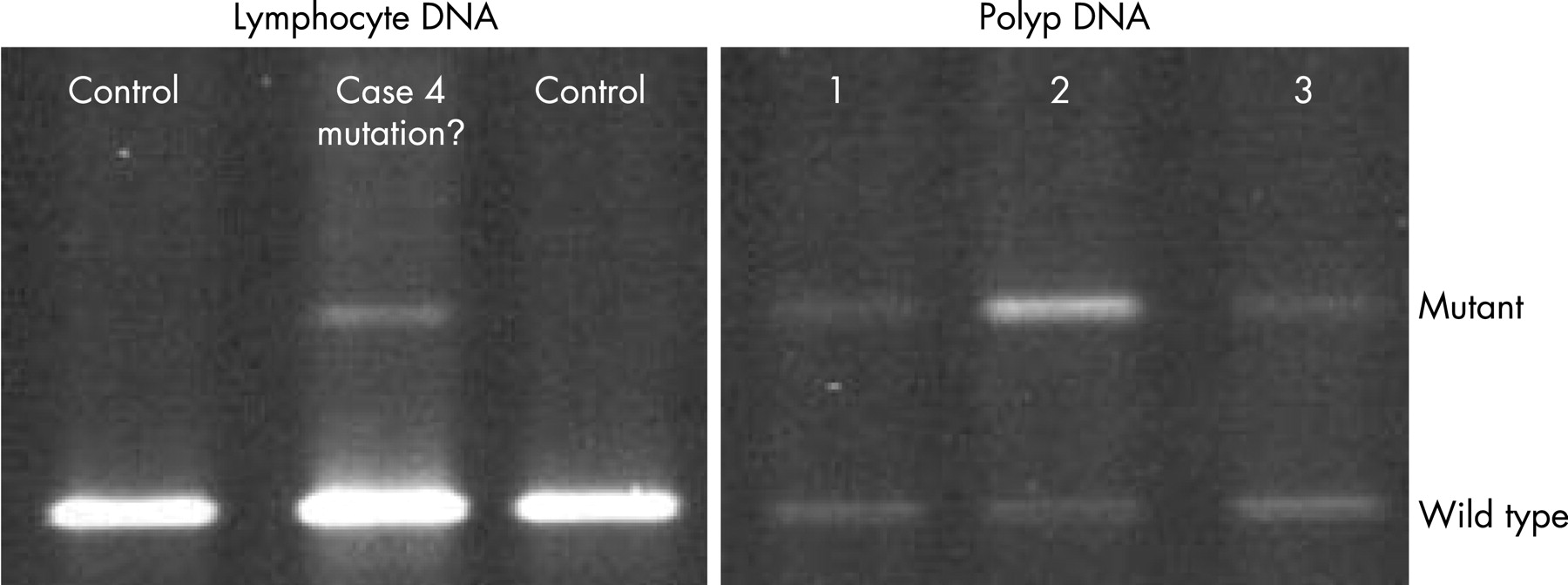
Five cases of somatic mosaicism (cases 1–5) were recognised in lymphocyte DNA by weaker signal intensity of the mutant bands as compared with the wild-type bands on DGGE (fig 1A). PTT showed a truncated band with low signal intensity in DNA from five adenomas of case 7 and in lymphocyte DNA of case 8. Two mosaic cases did show a close to 1:1 wild-type:mutant allele intensity on the PTT, but sequence analysis showed an excess of wild-type allele (cases 9 and 10). DNA derived from peripheral lymphocytes as well as from three adenomas was available in case 4; DGGE analysis showed the p.Arg216X mutation in both tissues, but by sequence analysis the mutant allele was not detectable in lymphocyte DNA (fig 2). In two patients (cases 5 and 6), the combination of clinical information and molecular genetic analysis indicated the presence of somatic mosaicism (fig 3).


Semi-quantification of the p.Arg283X mutation, observed in three mosaic cases, revealed that a fraction of 2–5% of mutated alleles could be detected using DGGE (fig 1A) and 10–15% with direct sequence analysis (fig 1B). In the total patient cohort (n = 599) the p.Arg283X mutation was found in two familial cases (with three generations) and four solitary cases. Three of these four solitary cases originated from a somatic (mitotic) mutation that resulted in a mosaicism. In a dilution PTT experiment, a fraction of 5% of the recurrent 5 bp deletion at codon 1309 could be visualised (data not shown). However, these observations were made after overexposure of the autoradiogram. Since, autoradiograms are not overexposed in daily use, the detection limit of mosaic bands using PTT would be in the order of 20%.
DISCUSSION
In many hereditary forms of cancer, such as colorectal (Lynch syndrome) and breast cancer, cases of mosaicism are probably not diagnosed due to referral bias. In these relatively frequently occurring forms of cancer, solitary patients are rarely referred for genetic counselling and/or testing. However, this situation is different for polyposis, since the diagnosis FAP can be made in a single person, and tens or hundreds of adenomas are very unlikely to occur by chance (i.e. many somatic mutations arising independently). In this study a systematic search for mosaicism was carried out in 242 families with germline APC mutations and resulted in the identification of 10 mosaic cases (4%). This relatively high number was not anticipated since only four cases of mosaic APC mutations have been reported so far.3–5 However, a high rate of mosaicism is likely in conditions with a high percentage of new mutations.6 The rate of new APC mutations has been estimated to be between 4×10⁻⁶ and 9×10⁻⁶ mutations per gamete per generation, and approximately 10–25% of all identified germline APC mutation carriers have a de novo mutation.15–17 Systematic studies in comparable (with respect to the proportion of de novo cases, mutation spectrum and frequency) tumour suppressor genes (RB1, TSC1 and TSC2) report mosaicism in approximately 10% of the identified germline mutations.10 11 25 26 Therefore, mosaic APC mutations have probably been overlooked before.
There are several ways in which mosaic mutations may remain undetected. First, the proportion of mutated cells could be under the detection level of the technique employed. In this study, DGGE and pyrosequencing detect fractions as low as 5% of mutated alleles. In other commonly used mutation-scanning techniques, such as direct nucleotide sequencing and PTT, the sensitivity is much lower (10–20%) and cases of mosaicism may well remain undetected. However, other techniques such as DHPLC (denaturing high-performance liquid chromatography), HRM (high resolution melt) analysis and PAP (pyrophosphorolysis-activated polymerisation) may be more sensitive and specific than our applied methods. Secondly, the number of cells with the mutation may be relatively high in peripheral blood and may be interpreted as a normal 1:1 ratio of mutant to wild-type alleles. Kehrer-Sawatzki et al. demonstrated that in NF1 patients carrying large deletions mosaicism, is frequently present.9 Remarkably, peripheral blood lymphocytes were found to have a higher proportion of mutated cells than other peripheral tissues, such as buccal smears or skin fibroblasts, suggesting that haematopoietic stem cells carrying an NF1 mutation may have a growth advantage over normal cells. Last, but not least, DNA isolated from peripheral blood leucocytes is most commonly used for mutation analysis, while mosaic mutations may be predominantly present in other cell lines, e.g. in the cells of the digestive tract (fig 2). This observation indicates that molecular genetic APC testing in sporadic polyposis patients should also use DNA retrieved from at least two independent adenomas.
Mutation type
Although the absolute number is admittedly small, we found significantly more C>T transitions in APC in cases with somatic mosaicism than in non-mosaic cases. Remarkably, all C>T transitions took place in CG sequences and, more specifically, in CGA sites, where a C>T transition leads to a Stop codon. This observation is in good agreement with the situation in haemophilia where C>T transitions in the factor VIII and IX genes occur more in families with somatic mosaicism than in those without, suggesting that CG sites might be more prone to early postzygotic mutations.27 28 Similarly, epidermal nevi arise from mosaic FGFR3 mutations that are almost exclusively C>T transitions at CG sites, whereas germline FGFR3 mutations cause autosomal dominant skeletal disorders such as achondroplasia and thanatophoric dysplasia, which can be associated with acanthosis nigricans of the skin.13
Genotype–phenotype correlations
Four of the somatic mosaics had a relatively mild polyposis phenotype (cases 3, 4, 7 and 10), whereas in the literature and from own observations the respective germline APC mutations (p.Arg216X, p.Arg283X and p.Thr1023fs) have been associated with florid forms of FAP.19 29 The germline mutation in case 8, p.Ser1436fs, has not been reported before to our knowledge, but is predicted to be associated with typical or even severe polyposis.30 31 We hypothesise that a relatively late mutational event could have consequences for a limited number of cells, e.g. part of the digestive tract, and thereby exerts a relatively mild phenotype. In FSHD, most mosaic mutation carriers are unaffected or very mildly affected, and the disease was only recognised in retrospect after symptomatic offspring were diagnosed.12
In contrast, five of our mosaic cases (cases 1, 2, 5, 8 and 9) displayed a more severe FAP phenotype including extracolonic manifestations. Both mosaic cases reported by Farrington and Dunlop also had a dense colonic polyposis at an early age, one of which had severe extracolonic manifestations.4 These florid manifestations of somatic mosaicsm might be explained by an early postzygotic event, involving different germ cell layers.
Finally, one case (case 6) was an apparently asymptomatic carrier of somatic mosaicism. Both sons, affected with thousands of adenomas, shared a maternally inherited APC-flanking haplotype. However, the germline mutation identified in the two sons could not be detected in lymphocyte and fibroblast DNA from the mother. This situation suggests somatic mosaicism in at least the gonadal cells of the mother. The 79-year-old mother was asymptomatic, but a colonoscopy was not performed because of her poor mental health. The father who developed colon polyps and carcinoma represents a phenocopy. Because of the misleading phenotype of the father, this family was earlier described as an APC-unlinked FAP family.23
Clinical consequences
The identification of mosaicism in a proportion of patients in which polyposis coli apparently arose de novo has consequences for clinical practice and genetic counselling (fig 4). Given the low but tangible number of occurrences of gonadal mosaicism and the possibility that low percentage mosaicism may remain undetected, we advise DNA testing to siblings of patients in which polyposis coli apparently arose de novo. An empirical recurrence risk of 7% in Duchenne muscular dystrophy,32 which is uncorrected for ascertainment bias, might also apply to polyposis coli. For offspring of carriers of a somatic mosaicism, the risk is ⩽50% depending on the level of mosaicism in the parental germ cells.33 34

As illustrated by case 5, somatic mosaicism might explain the occurrence of anticipation, defined as an earlier age of onset with a more severe phenotype in subsequent generations. Since regular mutation analysis might fail to detect somatic mosaicism, screening for germline APC mutations should preferably be conducted in affected children of de novo cases (i.e. with asymptomatic grandparents).
In conclusion, we have demonstrated a substantial role for mosaicism in polyposis patients, which frequently arises from mutations in CGA sites. Moreover, we anticipate identifying further mosaic cases in apparently sporadic patients without an identified germline mutation by using advanced mutation detection methods, by analysing tumour DNA in addition to lymphocyte DNA and focusing on CGA sites. Somatic mosaicism shows a wide phenotypic variability, probably depending on the timing and origin of the APC mutation. Mutations that appear de novo may in fact represent parental mosaicism and entail a recurrence risk for siblings. Finally, somatic mosaicism in patients with an attenuated phenotype may lead in their offspring to a more florid form of polyposis coli than expected.
Acknowledgments
The authors would like to thank Professor H. Morreau, pathologist, and Rolf Vassen for their valuable contributions.
REFERENCES
Footnotes
Funding: M.N. received a grant from the Dutch Digestive Diseases Foundation (grant MWO 0355). The funding agency had no input into the design and conduct of the study; collection, management, analysis and interpretation of the data; and preparation, review or approval of the manuscript.
Competing interests: None.