Article Text

Abstract
The novel Aristaless related homeobox gene, ARX, is widely expressed in the brain and is thought to play a key role in the regulation of brain development. Neurological phenotypes caused by ARX mutations have recently started to unfold. We describe a 72 year old man with X-linked mental retardation due to a 24 bp duplication mutation in exon 2 of the ARX gene. Cerebral MRI showed bilateral cystic-like cavities in both the cerebral and cerebellar hemispheres. No retraction or expansion in neighbouring parenchyma was observed, there was no history of acute neurological impairment, and no risk factors for cerebrovascular disease were found. The lesions appeared to be congenital and represented benign developmental cysts, possibly caused by the ARX mutation.
- ARX gene
- brain cysts
- X-linked mental retardation
Statistics from Altmetric.com
Congenital brain anomalies are not infrequently observed among mentally retarded individuals. In one population based study, structural brain anomalies were detected on neuroimaging in approximately 13% of individuals with prenatal aetiology behind mental retardation.1 If the aetiology is genetic, it is likely that the brain anomalies are associated with the same underlying genetic mechanisms. We describe a man who was an affected member of a family with X-linked mental retardation. Linkage analysis mapped the disease gene to the distal part of the short arm on chromosome X.2 The disease gene was subsequently identified to be the Aristaless related homeobox gene, ARX, localised in Xp22.1.3 The ARX gene belongs to the paired class of homeobox genes. These genes are transcription factors important in the regulation of brain development.4 Neurological features observed in the family in addition to mental retardation included infantile spasms, epilepsy, spasticity, and cerebellar ataxia.2 All affected individuals were investigated with neuroimaging studies. Bilateral cerebral and cerebellar cavities resembling benign congenital cysts were demonstrated in one patient.
CASE REPORT
The patient was a 72 year old man (fig 1) who lives in a home for the mentally handicapped. Little is known about his developmental milestones, although he has always been retarded and did not walk independently before the age of 5 years. He has only been hospitalised due to cholecystitis and pancreatitis. There have been no episodes of acute neurological impairment or seizures. Due to behavioural abnormalities, he was treated with pimozide 15mg daily. Chorea-like movements in his arms and neck were subsequently recorded. The medication was discontinued without any effect on the involuntary movements.
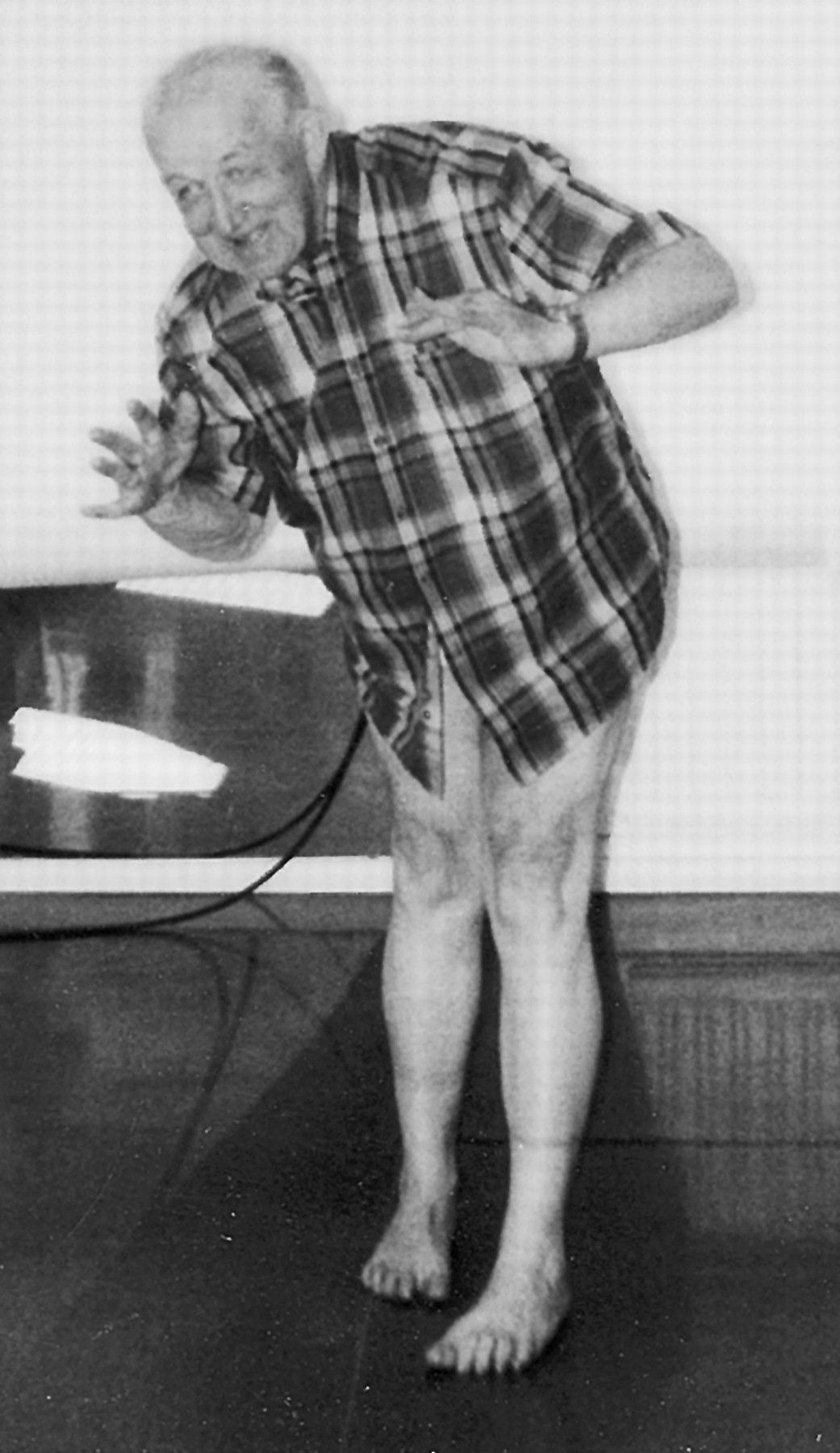
The patient demonstrating gait ataxia.
On examination he had a broad based spastic ataxic gait with symmetric and generalised hyperreflexia, without plantar inversion. Skilled movements of the hands were impaired. Dystonia involving both hands and facial muscles was noted. Cognitive functioning was in the range for the mildly retarded (IQ 50–70). Blood pressure, pulse, total cholesterol, blood glucose, Doppler ultrasound examination of the precerebral arteries, and an electroencephalogram were normal. Molecular genetic studies of the ARX gene demonstrated a 428–451dup (24bp) mutation in exon 2.3 The pathogenetic mechanism of this duplication, which results in addition of 8 extra alanine residues to the ARX protein, is currently not understood.
MRI examination showed fluid filled cavities in both cerebral (fig 2A) and cerebellar hemispheres (fig 2B). The lesions were located to the outer surface of the brain tissue. The left cerebellar lesion appeared to be confined from the subarachnoidal space by a cavity wall. The signal intensities on T1, T2, and proton density images, showed that the cavity fluid was cerebrospinal fluid (CSF). Further evaluation with intrathecal contrast medium and CT was not performed. There was no retraction or displacement of the surrounding parenchyma. The total volume of both the frontal lobes and the cerebral hemispheres were approximately equal. The corpus callosum was hypoplastic in its caudal part, while the basal ganglia and brain stem appeared normal. MRI or CT of other affected individuals were either normal or demonstrated hypoplasia of the cerebellum and corpus callosum.2 No other case with cavities was observed.

Proton density axial MR image of the cerebral hemispheres (A) demonstrating a large, partly CSF-filled cystic cavity in the left frontal lobe without communication to the ventricle. A similar, but smaller lesion (large arrow), is seen in the right frontal lobe. The margins of the lesions are sharply delineated and surrounded by gliosis (small arrows) as indicated by high signal intensity. The high signal changes along the lateral ventricles and the widening of the sulci in the parietooccipital region are normal findings in this age group. The rounded structure above the left ventricle (arrowhead) represents partly CSF and partly surrounding parenchyma, and was interpreted as partial volume effect. There are also CSF-filled cystic cavities in both cerebellar hemispheres towards the peripheries as shown on a T2 weighted image (B). In the left cerebellar hemisphere there is a rim of parenchyma (arrows) between the cyst and the subarachnoidal space. Both the carotid (c) and vertebral arteries (v) showed flow void indicating an open lumen.
DISCUSSION
Cerebral cystic lesions are unnatural cavities in which the continuity of the brain parenchyma is disrupted and replaced by fluid. Depending on the nature of the fluid content and the epithelial lining, cerebral cysts have been classified into the following categories: cysts containing CSF-like fluid, cysts with non-neural epithelium lining (colloid, epidermoid, and dermoid cysts), cysts associated with tumours, and infectious cysts.5 The mucoid protein and lipids rich contents of non-neural epithelium lined cysts give rise to bright T1 weighted MRI signals,6 not observed in the patient. Cysts associated with tumour or infection were unlikely considering lack of expansion and oedema in the surrounding tissues. Hence, the lesions in our patient were only compatible with the first category. This can be subdivided into three groups: ex vacuo type cysts (postraumatic leptomeningeal cysts, porencephalic cysts, or cysts resulting from surgical excision or vascular infarction), cysts with fluid secreting walls and CSF-like content (arachnoid and neuroepithelial cysts), and cysts associated with dysgenesis such as Dandy Walker and interhemispheric cysts. Porencephalic cysts frequently communicate with the ventricles and are associated with neurological deficits. Arachnoidal cysts are usually expansive and frequently located in the middle cranial fossa, while neuroepithelial cysts are formed within the ependymal lining of the ventricles. The dysgenesis group comprises heterogeneous developmental aetiologies, among which genetic aetiology should be considered.
As the cavities were lying at the outer margins, infarcts due to small vessel disease could be considered as a cause. However, the largest lesion in the left frontal lobe was too extensive to represent small vessel occlusion. Other features not favouring a vascular aetiology were lack of retraction in the surrounding parenchyma, and localisation of the lesions outside the watershed areas. Also, there was no history of acute neurological impairment, and he had no identifiable risk factors for cerebrovascular disease. Generalised hyperreflexia and cerebellar ataxia belonged to the phenotype associated with the mutation. However, the patient was not more spastic, ataxic, or mentally retarded than other affected individuals in the family. The lesions had therefore probably not contributed significantly to his motor or intellectual disabilities. The fact that he learned to walk independently at the age of 5 years suggested that the cause of his motor problems was congenital. Dystonia has also been observed in several other families affected by the same duplication mutation of the ARX gene. This particular mutation is associated with a wide range of neurological phenotypes.7,8
The most likely explanation for the cystic cavities was abnormalities of the developing foetal brain caused by the ARX mutation. The gene is widely expressed in the brain, including the telencephalon, ventral thalamus, cerebral cortex, amygdala, corpus callosum, caudate nucleus, and hippocampus.9 In a knock out mouse model Arx was recently shown to play an important role in neuronal proliferation and interneuronal migration and differentiation, as well as testicular differentiation. In humans, some ARX mutations were also shown to cause X-linked lissencephaly with ambiguous genitalia (XLAG).10 Mutations in the paired class of homeobox genes have been associated with malformations in the central nervous system, including the ocular region.11 Unilateral microphthalmia and structural brain asymmetry occurred in a boy with infantile spasms who had a deletion mutation of exon 5 of the ARX gene.3 The frequency of congenital cysts associated with mutations may not be high, although several affected individuals have not yet been studied with MRI. Interestingly, a large posterior fossa cyst was reported in one other case with an ARX mutation, a missense mutation of exon 2 associated with myoclonic epilepsy.12 Abnormalities in organs outside the central nervous system and genitourinary tract have not been reported.
In conclusion, it was most likely that the cystic cavities were related to the mutation in the ARX homeobox gene. The spectrum of phenotypes caused by mutations in this gene will probably expand as more cases become diagnosed.
Acknowledgments
This work was supported by The Unger-Vetlesen Medical Fund, Jersey, The Research Council of Norway and The National Health and Medical Research Council of Australia. We thank Rolf Nyberg-Hansen, Department of Neurology and Bengt Frode Kase, Department of Paediatric Research, Rikshospitalet, The National Hospital, Oslo, Norway, for review of the manuscript.