Article Text

Abstract
Familial atypical multiple mole melanoma syndrome (FAMMM) is characterised by dysplastic naevi, malignant melanoma and pancreatic cancer. Given that large deletions involving CDKN2A (cyclin-dependent kinase inhibitor 2A) account for only 2% of cases, we describe a family that highlights the co-occurrence of both melanoma and neural system tumours to aid clinical recognition and propose a management strategy. A patient with multiple neurofibromas was referred with a provisional diagnosis of neurofibromatosis type 1 (NF1). Prior molecular testing, though, had failed to identify an NF1 mutation by sequencing and multiplex ligation-dependent probe amplification. His family history was significant for multiple in situ/malignant melanomas at young ages and several different cancers reminiscent of an underlying syndrome. A search of the Familial Cancer Database, FaCD Online, highlighted several families with cutaneous melanoma and nervous system tumours who were subsequently identified to have large deletions spanning CDKN2A. Although sequencing of CDKN2A and TP53 failed to identify a mutation, a heterozygous CDKN2A deletion was identified by targeted array comparative genomic hybridisation (CGH). Whole-genome oligonucleotide array CGH and SNP analysis identified an interstitial deletion of at least 1.5 Mb within 9p21.3 and spanning approximately 25 genes. Identification of the underlying molecular abnormality permits predictive testing for at-risk relatives. Given the young cancer diagnoses, a surveillance regimen was developed and a clinical team organised for ongoing management so that genetic testing could be offered to both adults and minor children. Surveillance recommendations addressed cancer risks associated with FAMMM, and other cancers exhibited by this family with a large contiguous gene deletion.
- Cancer: CNS
- Cancer: dermatological
- Clinical genetics
- Genetic screening/counselling
- Molecular genetics
Statistics from Altmetric.com
Introduction
Familial atypical multiple mole melanoma syndrome (FAMMM) is an autosomal dominant cancer predisposition syndrome characterised by multiple dysplastic naevi, malignant melanoma and, in some families, a potential increased risk for pancreatic cancer. This cancer predisposition syndrome has also been referred to as dysplastic naevus syndrome and represents one of several genetic aetiologies underlying familial cutaneous malignant melanoma (FCMM).
The CDKN2A (cyclin-dependent kinase inhibitor 2A) gene, localised to chromosome 9p21, is the major known high-risk susceptibility gene for FAMMM/FCMM. CDKN2A codes for two distinct tumour suppressor proteins, p16INK4A and p14ARF, which are transcribed using alternative first exons, 1α and 1β, and subsequently spliced onto the common exons 2 and 3, but in different reading frames. p16INK4A acts through the pRb pathway and functions normally to inhibit the kinase activity of CDK4. In contrast, p14ARF exerts its biological effects through the p53 pathway. Ultimately, mutations in the CDKN2A gene can cause loss of function of either or both proteins, and so each may contribute to the development of different types of cancer.1
Germline CDKN2A mutations have been identified in upwards of 15%–40% of patients with FCMM, while an additional 1%–2% of familial cases are due to mutations in the CDK4 gene.2 ,3 Recently, three additional high-risk susceptibility genes for familial melanoma were identified, specifically BAP1, TERT and POT1, all of which are less frequent than CDK4.4–7 As such, there are likely other genes, not yet identified, that account for the remaining families with a hereditary predisposition to melanoma.
CDKN2A mutations were first reported in kindreds with familial melanoma in 1994 with missense mutations representing the predominant type of mutation identified.8 ,9 Nonsense mutations, as well as small deletions and insertions, have also been reported. Large deletions, though, involving one or more exons, account for only 2% of all mutations reported in the CDKN2A gene.3 ,10
Of the small number of families worldwide that have been described with large deletions involving CDKN2A, some have exhibited a predisposition to both melanoma and nervous system tumours (NST), prompting several investigators to propose that this combination of tumours may represent a discrete syndrome.11–14 Several case reports have specifically described these families as demonstrating tumours characteristic of both FAMMM and neurofibromatosis type 1 (NF1).14–17 For example, the family described by Bahuau et al14 exhibited many tumours associated with NF1, including both neurofibroma and astrocytoma, as well as features characteristic of FAMMM such as multiple cutaneous malignant melanoma and dysplastic naevi. The family reported by Bahuau et al, though, also exhibited some tumours characteristic of neurofibromatosis type 2 (NF2) such as schwannomas and meningiomas. We report a family with a large, contiguous gene deletion involving chromosome 9p21.3, and extending beyond CDKN2A to include approximately 25 genes, with tumours characteristic of FAMMM and NF1, and several additional tumours including a primitive neuroectodermal tumour (PNET), chondrosarcoma and leukaemia. The constellation of tumours exhibited by this family necessitated the development of a tailored surveillance regimen for ongoing clinical management of both adults, as well as minor children, given the high prevalence of young cancer diagnoses across multiple generations.
Methods
Patient and family
A 52-year-old Caucasian male with a provisional diagnosis of NF1 was referred to the Penn State Hershey Cancer Genetics Program by his oncologist for cancer genetic counselling and testing. A review of his personal history was significant for a squamous papilloma below the left eye which was diagnosed at 40 years of age, a benign fibrous histiocytoma of the lower back at 43 years of age, and one junctional naevus of the right upper arm with moderate to focal severe atypia at 44 years of age. In addition, his personal history was significant for multiple, painful neurofibromas which were diagnosed at 49 years of age, consisting of two in the retroperitoneum and one excised from the sciatic nerve. Subsequent review of his electronic medical record revealed that he had previously pursued a clinical genetics consult which included testing for NF1. The NF1 gene was analysed by long-range RT-PCR and sequencing using dye-terminator chemistry on an ABI PRISM capillary sequencer followed by multiplex ligation-dependent probe amplification (MLPA), the results of which were negative. Physical examination by a clinical geneticist documented the presence of multiple hyperpigmented naevi, but there was no evidence, besides the multiple nerve sheath tumours/neurofibromas, of other stigmata characteristic of NF1, such as Lisch nodules or café au lait spots. As such, the proband did not meet clinical diagnostic criteria for NF1.
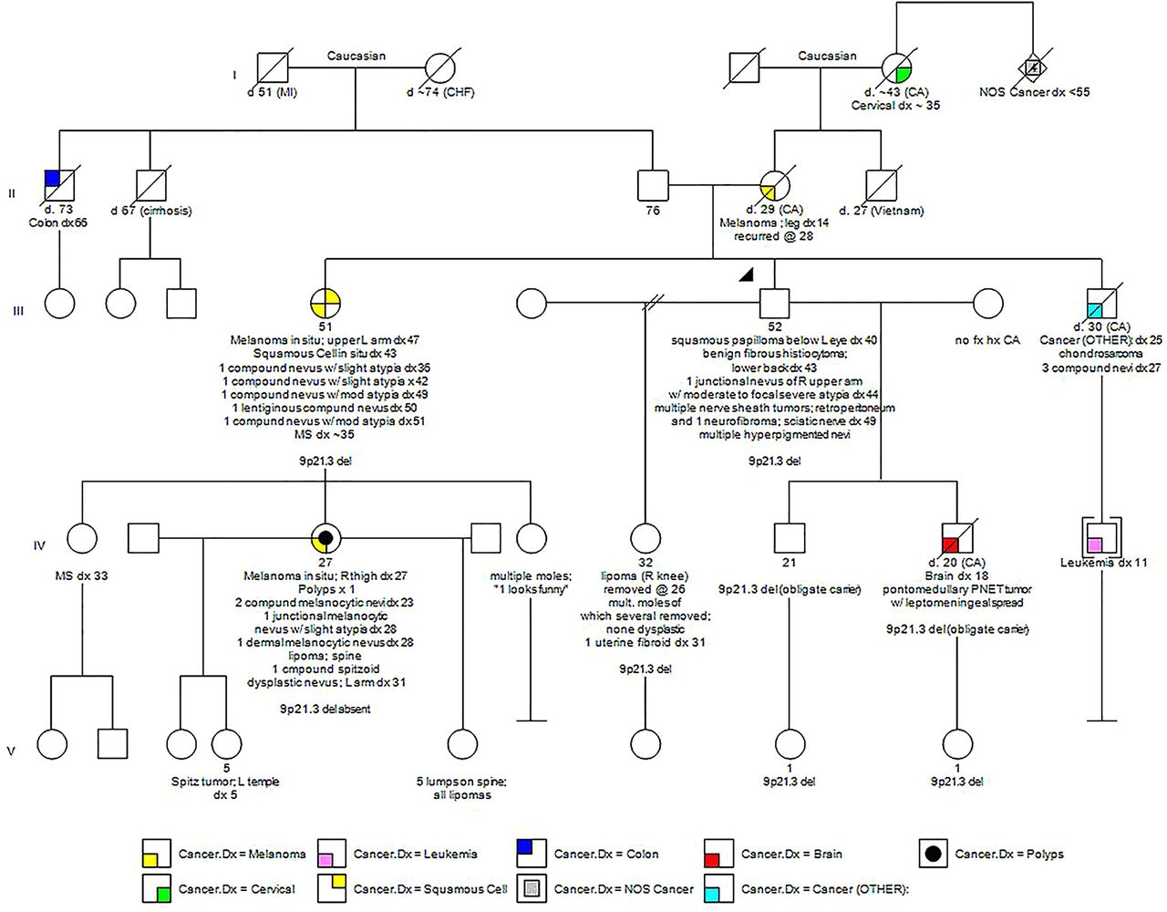
A review of his family history was significant for multiple relatives, spanning three generations, with both in situ and malignant cutaneous melanomas. His mother was diagnosed with a malignant melanoma at 14 years of age following which she died from metastatic disease at 29 years of age. In addition, his sister was diagnosed with melanoma in situ at 47 years of age and had a history of multiple naevi with atypia. The family history was also significant for a niece with melanoma in situ at 27 years of age. This niece had four additional naevi removed, one of which demonstrated slight atypia, and she had one lipoma. Of this niece's three daughters, one had a Spitz tumour excised at 5 years of age, possibly representing a precursor lesion to melanoma, and another daughter had five lipomas. In addition, the proband's family history was significant for his son who was diagnosed with a pontomedullary PNET with leptomeningeal spread at 18 years of age, his brother with chondrosarcoma of the inguinal region at 25 years of age followed by the removal of three compound naevi at 27 years of age and his brother's son with leukaemia which was diagnosed at 11 years of age. Other relatives with a history of cancer included his maternal grandmother with a reported diagnosis of cervical cancer at approximately 35 years of age and four great aunts and uncles, all with unspecified cancers under 55 years of age (see figure 1).

Family pedigree.
Genetic testing
Given that prior analysis of the NF1 gene was negative by both sequencing and MLPA, the proband was offered testing of the CDKN2A gene, which, at the time, consisted of sequencing only, to address the family history of multiple in situ and malignant melanomas and multiple atypical naevi. Amplified DNA products were sequenced in forward and reverse directions using fluorescent dye-labelled sequencing primers with chromatographic tracings of each amplicon analysed by a proprietary computer-based review followed by visual inspection and confirmation. In addition, he was offered both sequencing and MLPA analysis of the TP53 gene based on his family history of the PNET, chondrosarcoma and leukaemia, with mutations in this gene responsible for Li–Fraumeni syndrome. Amplified DNA was analysed by direct DNA sequence analysis on an automated fluorescent sequencer with sequencing of the entire coding region and associated splice junctions performed in both directions. MLPA products were analysed by DNA fragment analysis on an automated fluorescence sequencer with the absence or presence of deletions/duplications of one or more exons confirmed by MLPA analysis using an independently amplified segment. A search of the Familial Cancer Database, FaCD Online, revealed the identification of a small number of reported families worldwide with cutaneous malignant melanoma in the presence of NSTs, features reminiscent of both FAMMM and NF1, in the context of large deletions within and extending beyond CDKN2A that would not be detected by standard sequencing methods.18 ,19 As a result, the proband was also offered large rearrangement analysis of the CDKN2A gene via array-based comparative genomic hybridisation (aCGH), with the array containing multiple oligonucleotide probes in most exons and/or their flanking intronic regions, through another reference laboratory, if Sanger sequencing proved negative. Hybridisation data were analysed with the Genomic Workbench V.5 software (Agilent Technologies) to evaluate copy number at the exon level. Targeted exon-level array CGH was followed by whole-genome oligonucleotide array CGH and SNP analysis with the array design based on human genome build GRCh37/hg19.
Results
Genomic DNA was isolated from the proband's peripheral blood specimen. Subsequent sequencing, though, of the CDKN2A gene, including all exons and adjacent intronic regions, failed to identify a deleterious mutation underlying his personal and family history of cancer. Genetic testing also proved negative for a possible TP53 mutation following both sequencing and MLPA analysis. Targeted array CGH of the CDKN2A gene with exon-level resolution revealed a heterozygous deletion. Although this molecular result was informative for FCMM, the extent of the deletion beyond CDKN2A could not be determined by this assay alone. Subsequent whole-genome oligonucleotide array CGH and SNP analysis identified an interstitial deletion of at least 1.5 Mb within cytogenetic band 9p21.3 with sequence coordinates of chr9:20,951,885-22,447,709[hg19]. The deleted interval was found to include approximately 25 genes, of which only one, CDKN2A, was known to be associated with a cancer predisposition syndrome, specifically FCMM. See figure 2A,B. Additionally, the proband was identified to have two CNVs of unknown clinical significance: a duplication of at least 229 kb within cytogenetic band 5q35.2 with the duplicated interval including the TSPAN17 and EIF4E1B genes and part of the SNCB and UNC5A genes. The clinical consequence of carrying three copies of the SNCB gene, of which missense mutations have been described in two unrelated patients with dementia and lewy bodies, or of any of the other genes in this duplicated region has not yet been determined.11 Further, this region has not been reported to vary in copy number in the normal population.20 Lastly, the proband was found to have an amplification, consisting of four copies of at least 505 kb, in 10q24.32-q24.33. The amplified interval included 10 genes, none of which have been associated with clinical disorders to date.11 This region in its entirety has not been reported to vary in copy number in the normal population.20 In summary, the reference laboratory reported the molecular results as arr 5q35.2(176,052,444-176,281,813)x3,9p21.3(20,951,885-22,447,709)x1,10q24.32q24.33(104,853,173-105,357,653)x4 sex: male.

Array comparative genomic hybridisation (CGH) data. (A) shows the location of the deletion within cytogenetic band 9p21.3. (B) details the approximate 25 genes encompassed within the deleted region as identified in the proband. The green dots represent individual probe locations that are deleted in the proband compared with the reference DNA, based on the relative intensity of the signal. The probe locations are mapped in comparison with the genes in the genomic region. Red dots indicate probe locations whose intensity is increased in the proband relative to the reference DNA. Single probe deviations, whether red or green, represent hybridisation noise. Images provided by GeneDx.
Additional relatives tested to date include the proband's sister with multiple primary melanomas, an unaffected daughter and two unaffected granddaughters. Each of these relatives tested positive for the large interstitial deletion and, in the process, confirmed that both of the proband's sons were obligate carriers, one of whom was diagnosed with a pontomedullary PNET at 18 years of age. Most recently, the proband's niece with multiple primary melanomas tested negative for the contiguous gene deletion. This observation adds to the complexity of the family and raises the possibility that the constellation of cancers may result from multiple underlying genetic causes. Alternatively, the niece with melanoma, as well as other relatives, may be at increased risk for melanoma due to the presence of other familial risk factors.
Discussion
We report a family with a large deletion of chromosome 9p21.3, which spans approximately 25 genes and includes CDKN2A. To date, large rearrangement analysis of the CDKN2A gene has not routinely been offered by clinical reference laboratories when CDKN2A is ordered as a standalone test, since large deletions represent only 2% of CDKN2A mutations.3 ,10 Further, the proband presented for evaluation prior to the clinical availability of multiplex panels in which multiple cancer predisposition genes are analysed by next-generation sequencing, thus routinely permitting the detection of large rearrangements. The molecular aetiology underlying this family's history of tumours was aided by the availability of an online resource called the Familial Cancer Database, which is accessible at http://www.facd.info, and was primarily developed as a tool to assist healthcare providers in developing a differential diagnosis based on the constellation of tumours and non-tumour features within a family.18 ,19
CDKN2A-associated cancer spectrum
The identification of a large deletion encompassing CDKN2A confirmed a genetic predisposition to melanoma in the proband and a number of his at-risk relatives. In the context of FAMMM/FCMM, lifetime risk estimates for melanoma vary widely, with penetrance estimates ranging from a low of 28% by 80 years of age to a high of 58%–92%, depending on the study design.21–25 The Melanoma Genetics Consortium, GenoMEL, also found a statistically significant effect when families lived in a geographical area with a high population incidence of melanoma.26 They concluded that the risk factors which influence the population incidence of melanoma may also mediate the penetrance of CDKN2A mutations.
Pancreatic cancer has also been associated with mutations in the CDKN2A gene with one study estimating a 17% risk to age 75.27–29 Not all families, though, with a CDKN2A mutation demonstrate an increased risk of pancreatic cancer. Although previous studies have suggested that the development of pancreatic cancer may depend on whether the specific mutation identified impairs the function of the p14ARF protein in addition to p16, definitive evidence for this relationship has not yet been shown.
The melanoma-astrocytoma syndrome, first described in 1993, represents another phenotype postulated to be related to mutations in the CDKN2A gene.12 Since then, additional studies of families with melanoma have documented the co-occurrence of various neural system tumours, as seen in the proband's family, including rare solitary internal neurofibromas, as well as cutaneous neurofibromas.13 ,30 Lynch et al31 also documented the association of sarcoma with malignant melanoma in two kindreds with a CDKN2A mutation. Again, however, it has not yet been possible to clearly determine the underlying cause(s) of these rarely co-occurring tumours.
Genotype/phenotype correlations with large CDKN2A deletions
Although large germline deletions of CDKN2A have only been described in a limited number of families worldwide, the breakpoints of the deletions described thus far and their impact on the function of the gene's two alternative transcripts, p16INK4A and p14ARF, have begun to shed light on the underlying mechanism predisposing to the observed constellation of tumours, including dysplastic naevi, melanoma and neural system tumours. The first reported individual to carry a large deletion involving part of chromosome 9p, as a result of an unbalanced chromosomal translocation, developed multiple melanomas and a plexiform neurofibroma.16 It was estimated that the deletion in this patient spanned at least 6 Mb and involved CDKN2A, ARF and CDKN2B. Several years later, Bahuau et al32 reported two families with melanoma and various neural system tumours, including astrocytoma, meningioma, schwanomma and neurofibroma, both of which exhibited deletions of a portion of chromosome 9p.
Petronzelli et al proposed that p14ARF was responsible, at least in part, for predisposition to neural system tumours and melanoma based on a family that carried a germline splicing mutation that resulted in a lack of exon 2 sequences, thus rendering both proteins defective. They subsequently concluded that the development of neurofibromas was due to the inactivation of p16INK4A and p14ARF or, alternatively, of p14ARF alone.17 Pasmant et al33 detected a large germline deletion, which included the entire p15/CDKN2B-p16/CDKN2A-p14/ARF gene cluster, in a family with cutaneous malignant melanoma and neural system tumours, suggesting a contiguous gene deletion syndrome. However, in their study, they also identified a new long non-coding RNA, within the germline deletion, which they called ANRIL (Antisense Noncoding RNA in the INK4A Locus). More recently, Vanneste et al34 reported a patient with multiple neurofibromas and a solitary spinal neurofibroma who was found to have a deletion of 14 nucleotides in exon 2 of CDKN2A, providing further evidence that p14ARF, p16INK4A and/or ANRIL, now designated CDKN2B-AS1, may be specifically involved in the aetiology of neurofibromas as a feature of FAMMM. However, numerous FCMM families with large deletions impacting p14ARF do not have neural system tumours3 ,35 [Goldstein et al unpublished data 2015]. Thus, further study is required to understand the relationship between CMM, NST, CDKN2A, CDKN2B, CDKN2B-AS1 and other 9p21 genes. Although the contribution of ANRIL expression to neural system tumours remains unknown, SNPs which alter its expression, have been associated with numerous diseases including coronary artery disease, stroke, diabetes as well as melanoma and glioma.36 Most recently, Frigerio et al37 reported a patient with both astrocytoma and multiple melanomas with the largest constitutive deletion described to date involving 9p21.3 and spanning approximately 2135 Mb. Our proband, reported herein, adds to this growing list of families with large deletions extending beyond CDKN2A, thus raising the question of whether or not this could represent an emerging contiguous gene deletion syndrome.
Clinical implications
Based on the identification of a molecular deletion encompassing CDKN2A, the proband and his at-risk family members were instructed to follow FAMMM/FCMM surveillance recommendations which typically include total body skin examinations every 6–12 months by a dermatologist, beginning at 10 years of age and including whole body photography.24 ,38 Given the early-onset skin lesions within the family, though, baseline dermatological examinations were recommended to begin during the first few years of life. Most recently, the proband's niece with a history of multiple primary melanomas was identified not to carry the large deletion. Given that relatives who test negative for the known CDKN2A mutation remain at increased risk for melanoma due to other familial shared risk factors, though, they should pursue heightened skin surveillance, regardless of their genetic status.39 ,40 Lastly, with regard to dermatological recommendations, family members were educated regarding proper sun protection measures. Current FAMMM/FCMM surveillance recommendations also address the potential increased risk for pancreatic cancer in mutation positive family members. As such, the patient and his at-risk family members were instructed to discuss the role of endoscopic ultrasound of the pancreas, as well as possible measurement of the CA-19-9 tumour marker with a gastroenterologist.
Developing a tailored management strategy for this family, which addressed other cancer risks potentially associated with the large deletion, was limited by the fact that several relatives were unavailable for study. As a result, it was not possible to determine whether the brother and the nephew with respective diagnoses of chondrosarcoma and leukaemia each carried the large familial deletion. In contrast, the proband's son with the PNET, although unavailable for study, was determined to be an obligate carrier, given that his daughter tested positive for the familial deletion. As such, the following medical management recommendations were developed based on the family's specific history of benign tumours/cancers: (1) annual comprehensive physical examination, including a careful neurological examination, (2) consideration of whole-body MRI, (3) abdominal ultrasound and brain MRI on an annual basis and (4) bloodwork every 4 months to include complete blood count, erythrocyte sedimentation rate and lactate dehydrogenase. Given the proband's son who was diagnosed with a PNET at 18 years of age, baseline brain MRI was recommended beginning at 8 years of age. In addition, annual dilated ophthalmology evaluation by an ophthalmologist was recommended to look for optic glioma or papilledema, a sign of increased intracranial pressure which can occur secondary to the presence of a brain tumour. In summary, the medical team acknowledged that the proposed surveillance regimen was quite intensive. Given the proband's son, though, who, at the time, was actively dying from leptomeningeal spread of his PNET and the rather young diagnoses of melanoma within the family, this regimen was developed as a starting point for discussion with the clinical geneticist who ultimately would be responsible for overseeing the family's ongoing medical management. Lastly, although our proband did not meet clinical diagnostic criteria for NF1, nor did genetic testing confirm a molecular diagnosis, there could, likewise, be a potential risk to develop malignant peripheral nerve sheath tumours within the field of radiation treatment. As such, it was recommended that any decisions regarding radiation therapy be made in the context of a discussion with a radiation oncologist regarding the risks, benefits, and limitations of such treatment.41 ,42
Beyond the challenge of developing a management strategy for the proband and his at-risk relatives, given the paucity of families worldwide with a similar contiguous gene deletion, there was the ethical dilemma of whether presymptomatic testing should be offered to minor children. Typically, the appropriateness of offering presymptomatic testing to minors depends on the specific cancer predisposition syndrome segregating within the family and whether it is known to predispose to childhood cancers, and the phenotypic variability observed within the family. Given the early-onset cancers exhibited by family members and the development of an intensive management strategy, a number of the proband's relatives, both affected and unaffected, have since requested testing for both themselves and their minor children. To date, five additional relatives, ranging in age from 1 to 52 years of age, have now pursued testing and all but one was confirmed to carry the familial deletion.
Lastly, the proband's large deletion within 9p21.3 contains a number of candidate genes which make up the interferon gene cluster and have been shown to be important for patient survival and success of interferon therapy, beyond containing genes important in melanoma susceptibility.43 Linsley et al,44 for example, linked loss of this locus with reduced immune cell genes within melanoma tumours. They concluded that loss of 9p21.3 may lead or contribute to reduced immune surveillance and/or tumour destruction by the immune system. Thus, the family members described here, who develop melanoma in the context of the large deletion containing the interferon gene cluster, as well as others similarly affected with loss of 9p21.3, may be more likely to suffer metastatic disease and hence a worse prognosis. Chromosomal instability was recently demonstrated to be a mechanism for modulating local cytokine expression in colorectal tumours.45 Thus, emerging evidence suggests that genomic rearrangements within tumours may represent a broader mechanism for modulating antitumour immunity and, as such, could potentially influence the choice of treatment regimen in a family, such as the one presented here with a large deletion within 9p21.3, if and when tumours develop.44
Limitations
Our understanding regarding the spectrum of cancers associated with this family's contiguous gene deletion is limited by the paucity of families described in the literature with similar deletions. Further, within this family, it has yet to be determined whether the respective diagnoses of chondrosarcoma and leukaemia of brother and nephew occurred in conjunction with the familial deletion, whether there is more than one condition segregating within this family, or whether these diagnoses represent sporadic cancers within a family that has a genetic predisposition. For example, some of the features exhibited by the proband and/or his relatives are described in patients with PTEN hamartoma tumour syndrome (PHTS) such as the lipomas, the papilloma and the increased risk for melanoma. A next-generation sequencing panel, including PTEN, and potentially other cancer susceptibility genes, could address the possibility of an additional cancer syndrome segregating within the family. In addition, the specific contribution of the other genes within the contiguous deletion, if any, on the phenotype has not been explored. Lastly, although subsequent testing of the proband's sister identified that she shared both CNVs of unknown significance in common with her brother, in addition to the large deletion of chromosome 9p21.3, it has yet to be determined from which side(s) of the family the CNVs originated.
Conclusion
The family described here has a rare contiguous gene deletion which includes CDKN2A, and predisposes to multiple melanoma/dysplastic naevi characteristic of FAMMM/FCMM. The constellation of additional tumours within this family raises the question as to whether there is another cancer syndrome cosegregating within the family. The identification of the contiguous gene deletion underlying this family's hereditary predisposition to cancer was aided by the Familial Cancer Database which is a useful online tool to assist clinicians in the development of a differential diagnosis based on a family's specific history of various benign and malignant tumours. Based on our experience with this family and our review of similar cases within the literature, large rearrangement analysis of the CDKN2A gene should be considered if traditional Sanger sequencing of CDKN2A proves negative, when there is a family history of multiple melanoma concerning for FAMMM/FCMM, in the context of neural system tumours. Likewise, clinicians should consider both sequencing and large rearrangement analysis of the CDKN2A gene in patients, with neural system tumours suspicious, but not diagnostic for NF1 whose testing proves negative when there is a personal or family history of melanoma. Improved identification of these families will be further augmented by the increasing usage of next-generation sequencing pan cancer panels, which include CDKN2A, as well as whole-genome sequencing, both of which routinely detect large deletions and duplications, thus permitting better characterisation of the phenotype associated with this family's contiguous gene deletion. Lastly, predictive testing of minor children may be warranted in families with large deletions spanning CDKN2A, given the young cancer diagnoses observed in this family and assuming a clinical team can be assembled to develop a surveillance regimen that has the potential to impact prognosis of affected relatives. Further study of additional families with similar deletions spanning CDKN2A and beyond are needed to help guide genetic counselling and anticipatory care for these patients and to better understand this potentially evolving cancer predisposition syndrome.
Acknowledgments
The authors thank Kathleen Hruska, PhD, FACMG with GeneDx of Gaithersburg, MD for providing images illustrating the extent of the deleted region in the proband.
References
Footnotes
Contributors Genetic counselling and coordination of genetic testing: MJB. Ongoing medical management: PLG, KSH, HM, MJG and JJD. Drafting and critical revision of the manuscript: MJB, AMG, PLG, KSH, HM, MJG and JJD. All authors read and approved the final manuscript. KSH provided images of array CGH data.
Funding This work was supported in part by the Intramural Research Program of the US National Institutes of Health (NIH), National Cancer Institute (NCI), Division of Cancer Epidemiology and Genetics (DCEG).
Competing interests None declared.
Patient consent Obtained.
Provenance and peer review Not commissioned; externally peer reviewed.