Article Text

Abstract
The cardiofaciocutaneous (CFC) syndrome is a condition of sporadic occurrence, with patients showing multiple congenital anomalies and mental retardation. It is characterised by failure to thrive, relative macrocephaly, a distinctive face with prominent forehead, bitemporal constriction, absence of eyebrows, hypertelorism, downward-slanting palpebral fissures often with epicanthic folds, depressed nasal root and a bulbous tip of the nose. The cutaneous involvement consists of dry, hyperkeratotic, scaly skin, sparse and curly hair, and cavernous haemangiomata. Most patients have a congenital heart defect, most commonly pulmonic stenosis and hypertrophic cardiomyopathy. The developmental delay usually is moderate to severe. The syndrome is caused by gain-of-function mutations in four different genes BRAF, KRAS, mitogen-activated protein/extracellular signal-regulated kinase MEK1 and MEK2, all belonging to the same RAS–extracellular signal-regulated kinase (ERK) pathway that regulates cell differentiation, proliferation and apoptosis. The CFC syndrome is a member of a family of syndromes that includes the Noonan and Costello syndromes, presenting with phenotypic similarities. Noonan syndrome is caused by mutations in the protein tyrosine phosphatase SHP-2 gene (PTPN11), with a few people having a mutation in KRAS. Costello syndrome is caused by mutations in HRAS. The protein products of these genes also belong to the RAS–ERK pathway. Thus, the clinical overlap of these three conditions, which often poses a problem of differential diagnosis, is explained by their pathogenetic relatedness.
- ASD, atrial septal defect
- CFC, cardiofaciocutaneous
- ERK, extracellular signal-regulated kinase
- PTPN11, protein-tyrosine phosphatase nonreceptor type II encoding Tyrosine Phosphate SHP2
- PVS, pulmonary valve stenosis
Statistics from Altmetric.com
- ASD, atrial septal defect
- CFC, cardiofaciocutaneous
- ERK, extracellular signal-regulated kinase
- PTPN11, protein-tyrosine phosphatase nonreceptor type II encoding Tyrosine Phosphate SHP2
- PVS, pulmonary valve stenosis
The cardiofaciocutaneous (CFC) syndrome (OMIM 115150) is a syndrome where patients have multiple congenital anomalies or mental retardation, failure to thrive, psychomotor delay, a characteristic face, congenital heart defects, and abnormalities of the skin, eyes, gastrointestinal tract and central nervous system. Occurrence is sporadic, with men and women equally affected. The syndrome was first described 20 years ago by Reynolds et al1 in eight children. Additional reports soon followed and, according to a recent review,2 about 59 patients have been reported, providing the basis for an accurate delineation of the phenotypic spectrum of the syndrome. Nevertheless, a question has lingered for many years whether CFC is a unique and separate condition, or a variant of the Noonan syndrome (OMIM 163950)3–9 or of the Costello syndrome (OMIM 218040).10 These three conditions share several manifestations (table 1)11–13 and “borderline” cases do exist, usually in infants, which defy a clear-cut diagnosis. A useful diagnostic approach was provided with the creation of a CFC index based on 82 clinical traits and their frequencies in the population with the CFC syndrome.14 However, matters changed radically only with the discovery of different genes whose mutations cause each one of these syndromes: the protein tyrosine phosphatase SHP-2 gene PTPN11 for Noonan syndrome,15HRAS for Costello syndrome,16 and KRAS, BRAF, mitogen-activated protein/extracellular signal-regulated kinase MEK1 and MEK2 for CFC.17,18 These discoveries are doubly interesting for (a) finally allowing clarification of the nosology of these three syndromes and (b) explaining their similarities, given that the protein products of the causative genes interact in a common pathway.18 Therefore, it seems timely and appropriate to review the status of the CFC syndrome, providing a thorough clinical and molecular description and a nosological discussion with respect to the Noonan and Costello syndromes (table 1).
Phenotypic overlap of the cardiofaciocutaneous, Noonan and Costello syndromes
PREVALENCE AND HERITABILITY
No existing neonatal screening or epidemiological studies provide an accurate estimate of the population prevalence of the CFC syndrome. As noted above, about 60 cases are published, most likely representing the tip of an iceberg. More than 100 unpublished cases are known to CFC International, a family-support group operating worldwide (http://www.cfcsyndrome.org). Even this number must be an underestimate, excluding most of the mildest cases. Molecular confirmation of clinically suspected cases will soon add valuable data to the prevalence issue. All cases described so far are sporadic, most likely due to new dominant mutations, as suggested by the observation of a paternal age effect.19
THE PHENOTYPE
Growth and longevity
Reynolds et al1 reported that pregnancies of affected women may be complicated by polyhydramnios, although measurements at birth tend to be normal. Postnatally, there is failure to thrive, psychomotor delay and generalised hypotonia.
Short stature is found in 78% of patients.20 Weight is also below the normal growth curve, but there is relative macrocephaly. Bone age is considerably delayed and osteopenia is occasionally observed.21 The typical progression is that a child is born of average weight but then there is failure to thrive, often due to feeding problems.
On the basis of prolonged follow-up of several affected individuals, it is our impression that the phenotype changes over the years—for example, the face becoming less typical and the hair becoming thicker and more normal. Little is known about the fertility of people with the CFC syndrome. No cases are known of children born to a bona fide parent with the CFC syndrome. Life expectancy is probably shortened on average, due to the early death of those with severe cardiac involvement.
Development and the neurological system
Neurological involvement in the CFC syndrome is extensive, and can involve functions of the cortex, brain stem and ventricular system. Mental retardation and global developmental delay are found in most (81%) cases.20 The less drastically affected children are probably not diagnosed as having the CFC syndrome. As molecular genetic testing is completed on more children, the developmental phenotype may broaden to include children with less severe, or little, developmental delay or mental retardation. When children are serially examined, there is a moderate to marked psychomotor delay in infancy and early childhood, which becomes less obvious with age.5 All cases describe delayed language acquisition. There is one case published of a 25-year-old woman with the CFC syndrome without mental retardation. She attended school up to age 19 years, with good results. No specific learning disabilities were reported and she was employed as an administrative assistant.22 Multiple structural brain abnormalities have been reported including cerebral atrophy,23–26 frontal lobe hypoplasia,1 absence or hypoplasia of the corpus callosum,10 increased ventricular size or hydrocephalus,1,24 non-specific leucodystrophy of the right frontal region,26 brain stem atrophy,10,27 widening of the cerebral sulci and the basal cisternae,28 and hypoplasia of the cerebellar vermis.10 Hyperactive deep-tendon reflexes have also been reported.1 Considerable hypotonia leads to marked motor delay. Some children have characteristic recurrent episodes of eyelid fluttering brought on by minimal stimulus.24 Seizures are reported but more often abnormalities can be found on an electroencephalogram, including decreased anterior voltage, spike-wave or polyspike pattern, sharp and slow waves, generalised dysrhythmia grade I or II, irritative waves, or generalised disorganisation, even when clinically apparent seizures are absent.1,25,26 One child was considered to have Lennox–Gastaux syndrome.29
The face
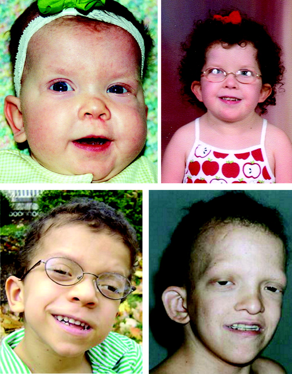
Children with the CFC syndrome have a relatively large head, a tall forehead with narrowing at the temples, palpebral ptosis and a short nose with a relatively broad nasal base. The philtrum has a deep groove with cupid’s bow lip and a small chin. The eyes are wide spaced and the palpebral fissures are often downward slanting, with epicanthic folds (fig 1). These findings are similar to those seen in children with Noonan syndrome, particularly those under 5 or 6 years of age. However, even in this age range, the face tends to be more “coarse” than that seen in case of the Noonan syndrome, and dolichocephaly is more likely to be present. The typical ear shape and placement in the Noonan syndrome, oval with an over-folded helix, low set and posteriorly angulated, is uncommon in the CFC syndrome. Earlobe creases appear quite frequent. At older ages, the face is broad and coarse, and lacks the inverted triangular shape seen in the Noonan syndrome. Iris colour is rarely the characteristic blue or blue–green as seen in the Noonan syndrome. There is a high likelihood of absent eyebrows with hyperkeratosis (ulerythema ophryogenes). The scalp hair is usually sparse, curly and friable.

Three patients of various ages with the cardiofaciocutaneous syndrome. The face is typical, with a broad forehead, bulbous tip of the nose, low-set ears, sparse scalp hair and absent eyebrows with ulerythema ophryogenes. The upper panel shows the same child at 10 months (left) and 6 years (right), describing the evolution of the phenotype. All of these children carry a BRAF mutation. Parents of patients gave written consent to publish these images.
Allanson et al30 carried out a detailed examination and measurement of 35 children with the CFC syndrome. Anthropometric assessment showed increased facial widths with facial depths and circumferences closer to normal, a broad nose and mouth, and wide-spaced eyes. In comparison with the Noonan syndrome, the face is both broader and longer.
Skin and adnexa
Cutaneous or adnexal abnormalities are seen in the CFC syndrome and in the phenotypically similar Noonan and Costello syndromes. Our observation of patients with the CFC and Costello syndromes (about 150), who attended family-support group conferences for both syndromes, has shown that skin or adnexal abnormalities are present in 100% of people affected. However, the clinical presentation is different and in some cases suggests the diagnosis.
Those with the CFC syndrome present with follicular hyperkeratosis of the arms, legs and face as the most common cutaneous abnormality (table 2; fig 2). Sparse, slow-growing, curly hair is another hallmark of the CFC syndrome, and is present in 85% of those affected (table 2). On the other hand, patients with the Costello syndrome show palmoplantar hyperkeratosis, skin tags and warts, and nail dystrophy as the most common skin and adnexal anomalies. The presentation of palmoplantar hyperkeratosis also differs from that seen in the CFC syndrome. In people with the Costello syndrome, it is found outside the pressure zones, whereas in those with the CFC syndrome, it is mainly present in the pressure zones.
Skin and adnexa findings in people with the cardiofaciocutaneous syndrome

Examples of cavernous haemangioma and ulerythema ophryogenes.
Only a few publications have dealt in depth with the cutaneous findings in the CFC syndrome. Histological findings31–33 were relatively non-specific, showing ichthyosis and hyperkeratosis of sweat glands and hair follicles.
Almost two decades of follow-up of some people with the CFC syndrome has shown that, with age, the dryness of the skin and the follicular hyperkeratosis tend to improve, allowing the hair to grow on the scalp and face. However, palmoplantar hyperkeratosis and lymphoedema may become more severe. Lymphoedema and its complications may have been the cause of death of a teenage boy (personal observation). Therefore, careful follow-up of patients presenting with lymphoedema and treatment of skin and nail infections is highly recommended.
We have seen no complications related to the pigmented naevi. Periodic evaluation of the pigmented lesions, as well as excision of these lesions in traumatised areas, is recommended, although we have not seen malignant transformations of such lesions.
The heart
Data on cardiac evaluations are available on 53 individuals (published and unpublished) with the CFC syndrome, including three with autopsy (table 3).34–39 Cardiac ultrasonography showed no evidence of abnormality in 13 (24.5%) people. The remaining 75.5% had one or more abnormality. Atrial septal defect (ASD), either isolated or associated with pulmonary valve stenosis (PVS), was noted in 9 (22.5%) of those with a cardiac abnormality. Five of these also had mild pulmonary stenosis, two had mild hypertrophic cardiomyopathy and one had severe hypertrophic cardiomyopathy. Surgical repair of the ASD was successful in three patients. One patient required mitral valve replacement because of prolapse with severe insufficiency. Another person underwent closure of an ASD with an Amplatz device. In 13 patients, PVS was the predominant lesion, and in five others it was associated with an ASD. Thus, 18 (45%) had PVS. It was mild or trivial in 15 patients. Two had a surgical pulmonary valvotomy and one had a balloon valvuloplasty. One patient, in addition to PVS, had mitral valve prolapse and another had mild hypertrophic cardiomyopathy. Other cardiac lesions included small ventricular septal defects in two patients and a partial atrioventricular canal in another two patients, one of whom has had successful repair. Another patient had a thickened mitral valve and another mitral valve prolapse. Twelve people had some form of myocardial disease as the primary diagnosis, which was associated with another defect in four others. Thus, 16 (40%) had some form of myocardial disease. Eight had mild hypertrophic cardiomyopathy, two had a localised bulge in the subaortic area and one child had moderate diastolic dysfunction. Two patients were receiving treatment because of major hypertrophic cardiomyopathy with obstruction. Another two patients with severe hypertrophic cardiomyopathy died suddenly at ages 21 and 22 years.
Overlapping heart involvement in cardiofaciocutaneous, Noonan and Costello syndromes
Autopsy findings were available in three affected patients. A 4-year-old girl died after a brief course of high fever, diarrhoea and a blue, painful right foot. Bacterial endocarditis due to Staphylococcus aureus was documented. Autopsy showed a pulmonary valvectomy carried out at 2 years and an ASD, 8 mm in diameter. All four valves were dysplastic. Extensive vegetations were present on the mitral and aortic valves, with multiple infarcts throughout the body. A 21-year-old man died suddenly. A murmur heard in early childhood was attributed to mild PVS. Hypertrophic cardiomyopathy was not recognised until shortly before his death. Autopsy documented biventricular and septal hypertrophy. The pulmonary valve was normal, but the mitral valve was thickened, and the aortic valve was dysplastic with irregular malformed leaflets. Microscopical examination showed the expected focal myofibril disarray, and the intramural coronary arteries showed intimal thickening and early fibrosis, consistent with hypertrophic cardiomyopathy. An unexpected finding was noted in the lungs. The pulmonary artery showed marked intimal thickening, with a narrow lumen consistent with grade 3 pulmonary hypertension. A 22-year-old man died shortly after the sudden onset of ventricular fibrillation. A diagnosis of the CFC syndrome had been made in childhood, and an ASD and a PVS had been successfully surgically repaired. At age 20 years, tachycardia and arrhythmia became untreatable problems. At age 22 years, he developed congestive cardiac failure. At autopsy, marked cardiomegaly with microscopic disarray of myocardial fibres, characteristic of hypertrophic cardiomyopathy, was observed.
Cardiac findings in the CFC syndrome are remarkably similar to those noted in the Noonan and Costello syndromes (table 3). Patients with the Noonan syndrome with a PTPN11 mutation have a considerably higher incidence of PVS but a lower incidence of hypertrophic cardiomyopathy.34 Unlike in the Noonan or CFC syndromes, atrial tachycardias or other arrhythmias occur in >30% of patients with the Costello syndrome, particularly in infancy.35,40 The incidence of PVS and hypertrophic cardiomyopathy are similar in CFC and Costello syndromes, whereas Noonan syndrome has a considerably higher incidence of PVS and a much lower incidence of hypertrophic cardiomyopathy. The LEOPARD syndrome, an allelic variant of the Noonan syndrome, has a high incidence of hypertrophic cardiomyopathy.36 The overall incidence of heart disease in the LEOPARD syndrome is 65%, and 80% of those with a cardiac abnormality have hypertrophic cardiomyopathy.41
The congenital defects in all three syndromes can be managed as in any child. However, little is known about the natural history of hypertrophic cardiomyopathy, whose course and prognosis are variable and not well understood. From the experience in the Noonan syndrome and from the limited reports on the CFC and Costello syndromes, marked variability is apparent in the natural history of hypertrophic cardiomyopathy. This condition may be rapidly progressive in infancy42 or may remain stable for many years.43 It may develop late in childhood, and it may resolve, remain stable or progress. Symptomatic hypertrophic cardiomyopathy in infancy is associated with considerable mortality. Although the risk of sudden death in the Noonan syndrome in asymptomatic patients is not well known, sudden unexpected death has been reported.44 The treatment for hypertrophic cardiomyopathy is similar to that in children without the syndrome. Some may improve with high-dose β blockers.45 Surgical relief of the symptomatic person with obstructive cardiomyopathy may be successful and an occasional patient has undergone cardiac transplantation.
Non-syndromic hypertrophic cardiomyopathy, often familial, is attributed to mutations in several genes, including the α-tropomyosin and cardiac troponin T genes.46 It is a heterogeneous disease of sarcomeric proteins and is an important cause of sudden unexpected death in young adults. The myocardial findings in patients with the CFC syndrome and related disorders are similarly characterised by myocardial disarray and thick-walled intramural coronary arteries.44,47 However, unlike the non-syndromic form, the right and the left ventricles are often involved.48
In conclusion, our knowledge about the long-term prognosis of the congenital heart defects in the CFC syndrome is still limited. It is clearly necessary to continue monitoring the cardiovascular system with increasing age. The finding of primary pulmonary hypertension is of interest. This condition has also been reported in several patients with the Noonan syndrome.
Bleeding disorders
Unlike the Noonan syndrome,49 easy bruising and bleeding problems have not been recognised as major problems in the CFC syndrome. Among the patients we have seen, one had transient thrombocytopenia as a newborn and another had frequent nosebleeds that improved with cautery. Easy bruising was not reported by any individual. Haematological data are not available.
The eye
Ocular manifestations are common in the CFC syndrome. Most of them show supraorbital ridge hypoplasia, hypertelorism, downward-slanting palpebral fissures, ptosis, nystagmus, strabismus or other abnormalities of the eye that require careful management by an ophthalmologist. To date, there is little information in the literature describing the ocular phenotype of this condition. To our knowledge, Young’s50 summary of the ocular findings of 12 patients with the CFC syndrome attending the CFC conference in Rockville, Maryland, USA, in 2003, and the report by Young et al51 describing the findings in the eye of 3 children with the CFC syndrome are the only published descriptions of ophthalmic findings in this condition. These results can now be confirmed by the observation of 13 additional individuals. Table 4 summarises the demographic characteristics and ophthalmic findings this group. Of the 13, six were male and seven were female. The ages ranged from birth to 26 years (mean 6.8 (standard deviation 6.6) years).
Demographics and data of people with the cardiofaciocutaneous syndrome undergoing an ophthalmological examination
One of the most notable findings in the previous and present studies was the considerable range of visual function among people with the CFC syndrome. Loss of vision to the level of light perception was rare, whereas decrease in visual acuity was more common. Strabismus was also common, affecting about one third of patients. An exotropic deviation was more common than esotropia. A higher percentage of children in the present group had nystagmus (7/13; 54%) than that described by Young et al51 (2/12; 17%). This study also shows a higher prevalence of myopia (4/13; 31%) compared with that in Young’s50 summary (1/12; 8%), although only two of four patients with myopia were prescribed spectacle correction.
The only optic nerve finding that occurred in more than one patient was optic nerve hypoplasia. One person had optic atrophy, suggesting a possible increase in intracranial pressure. There were only isolated cases of cataracts, vertical strabismus, dissociated vertical deviation and inferior oblique muscle overaction, nasolacrimal duct obstruction, ptosis and keratoconus. Ocular coloboma, lens dislocation or other structural changes in the eye were not observed. Most children evaluated had difficulty with depth perception and binocular function. Although this may be due to visual-processing challenges, an overlying strabismus or nystagmus can also contribute to fixation and tracking abnormalities. Studies on a larger number of patients are needed to confirm the ocular phenotype of the CFC syndrome.
In conclusion, it is important that children with the CFC syndrome undergo a comprehensive ophthalmological evaluation as early as possible in the newborn period to assess visual disturbances, including strabismus and refractive error, which may ultimately lead to amblyopia.
Gastrointestinal tract
Feeding problems are seen in the neonatal period, characterised by poor suck, aspiration, gastro-oesophageal reflux, oral aversion, hyperemesis and gastrointestinal dysmotility. These problems are important enough to often require nasogastric tube feeding, gastrostomy tube placement and/or Nissen fundoplication. Structural abnormalities have also been reported. There have been two cases with malrotation and antral foveolar hyperplasia.21 Antral foveolar hyperplasia is often an acquired abnormality caused by an inflammatory infiltrate, but cultures and stains were negative in the reported case and the cause remains uncertain.21 McDaniel and Fujimoto52 reported a child admitted for inadequate weight gain and frequent postprandial vomiting. Gastrointestinal evaluation showed malrotation of the small intestine, which was surgically corrected. Severe constipation (with normal ganglion cells) requiring disimpaction has also been reported.21 Nanda et al53 reported an unusual case of a 17-year-old young woman with the CFC syndrome, with a history of recurrent intermittent abdominal pain and fatty changes in the liver. Liver biopsy showed non-specific fatty changes (mixed macrovesicular and microvesicular steatosis). Ion et al27 reported one child with Crohn’s disease. The case of a toddler with food intolerance, especially to milk, fish and egg, with episodes of asthma, diarrhoea and vomiting was also reported.28 Additional digestive system findings include hepatomegaly, umbilical hernia, inguinal hernia, anal stenosis and intestinal malrotation.52
MOLECULAR ASPECTS
Since the original description in 1986, it has taken 20 years to discover genes whose mutations cause the CFC syndrome and to indisputably establish this condition as a distinct genetic entity, with autosomal dominant heritability. The nosological tangle involving the CFC, Noonan and Costello syndromes was partly solved by the discovery in 2001 of PTPN11 as a gene responsible for a large proportion of patients with the Noonan syndrome.15 Reports soon followed, showing that PTPN11 mutations are not found in those with a firm clinical diagnosis of the CFC syndrome.20,27 Further clarifying evidence was provided by the report in 2005 that the Costello syndrome is caused by HRAS mutations.16 These mutations were also shown not to be involved in the CFC syndrome.54
The final evidence came with the recent discovery that yet other genes cause the CFC syndrome. Rodriguez-Viciana et al,17 studying 23 individuals with the CFC syndrome, found 11 different mutations of BRAF in 18 of them, mutations of MEK1 in two and of MEK2 in one person. All of these were de novo mis-sense mutations, suggesting a gain-of-function effect. Likewise, Niihori et al,18 studying a non-overlapping group of 43 patients with the CFC syndrome, found eight different de novo mis-sense mutations in BRAF in 16 and also de novo mis-sense mutations in KRAS in three of them. All patients from Niihori et al’s18 group testing positive for any one of the reported mutations had a typical CFC phenotype (growth failure, mental retardation, relative macrocephaly, characteristic face, curly and sparse hair, heart defects). On the other hand, not all typical cases of the CFC syndrome have one of the known mutations. Out of 10 bona fide cases described by Kavamura et al,20 only five tested positive for a mutation (Dr Aoki, personal communication, 2006). With respect to genotype–phenotype correlations, Niihori et al18 found that skin abnormalities were present in BRAF-positive patients, but not in KRAS-positive ones. No other major differences were noted. By pooling data from the two reports (table 5), there are 13 different BRAF mutations, Q257R being the most common (n = 8).
Genes involved in the causation of the cardiofaciocutaneous, Noonan and Costello syndromes
The protein products of the CFC genes and those of HRAS involved in the causation of Costello syndrome,16 and those of PTPN11 in the Noonan syndrome15 (table 5), all have a role in the RAS—extracellular signal-regulated kinase (ERK) pathway (fig 3). RAS genes encode guanosine triphosphate-binding proteins that serve as molecular on–off switches that activate or inhibit downstream molecules. It is a signalling pathway that is important for cell proliferation, growth and death. When dysfunctional, it can cause cancer. A major proportion of mutations are gain-of-function mutations, as shown by in vitro assays, and stimulate the RAS–ERK pathway. This might explain the increased incidence of solid tumours (rhabdomyosarcoma, ganglioneuroblastoma, bladder carcinoma, etc) in patients with the Costello syndrome and of haematopoietic malignancies in those with the Noonan syndrome.62 Conversely, no increased incidence of tumours has been noted so far in patients with the CFC syndrome. It is not clear whether acute lymphoblastic leukaemia in one patient with a BRAF mutation18 should be considered to be a component manifestation of the syndrome or just a coincidence. However, increased cellular proliferation could explain some of the clinical findings in the CFC syndrome, such as hyperkeratosis and hypertrophic cardiomyopathy. Still, the major effect of all described mutations seems to take place during development, explaining psychomotor retardation and physical anomalies, the common denominator of all three syndromes.

RAS–extracellular signal-regulated kinase (ERK) signalling pathway connecting pathogenetically the cardiofaciocutaneous (CFC), Costello and Noonan syndromes. Inactive HRAS and KRAS (green outline) are activated (red outline) by neurofibromin and SHP2. Red arrowheads, activation; black arrowheads, inhibition.
Many important details need further clarification through the discovery of additional causative genes. For instance, a good proportion of patients with the Noonan syndrome do not have a PTPN11 mutation and, although a recent report established that KRAS mutations can also cause the Noonan syndrome, mutations were found in only a small proportion of cases. These individuals were described to have more severe features than would be expected in typical Noonan syndrome.57 The complexity of the issue is well considered by Bentires-Alj et al,63 who dissected the many components of the RAS pathway in relation to the Noonan, CFC, Costello and neurofibromatosis syndromes. At the moment, however, there are more questions than answers in trying to establish why pathogenetically related syndromes display major phenotypic differences.
Yet, we can firmly state that the CFC, Noonan and Costello syndromes are genetically heterogeneous. As they are all caused by mutations in genes whose protein products are part of the RAS–ERK pathway, we also understand, at least partly, why they are phenotypically similar. Further clarification may result from new molecular findings also, but perhaps more likely, and from a more detailed phenotypic description of the various affected patients.
Historical colophon
Virtually every well-defined condition or syndrome in medical genetics (ever more frequently causally identified) has a classic historical precedent as de Lange had in Brachmann, Étienne-Louis Arthur Fallot (tetratology) in Niels Stensen and Johann Friedrich Meckel the Younger (the latter fully aware of the resulting defect in oxygenation), Turner (45,X) in Ullrich and George Fraser in the German-speaking cryptophthalmos pioneers Zehender, Chiari and Fuchs64. The list is endless and the CFC syndrome is no exception.
First published synoptically, after some editorial surgery, upon evaluation of eight patients by Reynolds et al,1 it was preceded by at least one report by Navaratnam and Hodgson,65 who described the entity ulerythema ophryogenes in patients who obviously had what is now known as the CFC syndrome.
In the same year that saw the publication of Reynolds et al,1 Baraitser and Patton66 evidently described the same entity—another one of innumerable recent instances of “when the time is right” discoveries of the same condition or phenomenon, independently in one part of the world or another.
All biological entities have two histories: an ontogeny in a specific individual and a species-specific phylogeny involving a group of individuals. Again, the CFC syndrome is no exception. The ontogeny of the condition is encapsulated in the eight clinical reports by Reynolds et al1; however, that publication had a phylogeny spanning almost two decades.
The first child with the CFC syndrome studied by JMO was SVH (patient 3 in Reynolds et al1), born on 20 July 1968 and ascertained on 9 December 1968 at the former Wisconsin Orthopedic Hospital for Children of the University of Wisconsin, Wisconsin, Madison, USA after a visit to the cardiology clinic for probable pulmonic stenosis and atrial septal defect. He was followed up for some years, also studied by FR Grosse and Gerhard Neuhäuser, and then lost to follow-up. An effort has been initiated to determine his subsequent fate.
The second child from Wisconsin was KAS, patient 2 in Reynolds et al,1 born on 11 March 1969 and ascertained on 28 July 1969. She was one of two of the original three children with the CFC syndrome with a cavernous hemangioma. She had a heart failure presumably due to an endocardial cushion defect. Her present condition is unknown. Patient 1 in Reynolds et al1 was the third of the children with the CFC syndrome from Wisconsin, born on 5 July 1967 and ascertained on 19 April 1971, also with a hemangioma. An attempt to interest former Wisconsin coworkers in a collaborative publication at that time was unsuccessful.
About 10 years after the Wisconsin group began collaborating with Dr Philip D Pallister in Montana, we studied KL (patient 6 in Reynolds et al,1 born on 2 September 1974 and first seen by Dr Pallister on 17 December 1976). In west America, especially Montana, families tend to “stay put”; thus, it was no surprise that this young woman’s mother called promptly at 08:00 on 1 March 2006 from her original phone of 30 years ago, delighted at the news that no less than four genes had been identified as the cause of the CFC syndrome and to tell us that her daughter was in stable condition, doing well at age 31 years. She had graduated from high school in special education and was now living, self-sufficiently in an apartment space of her own in the family’s original home with her sister, brother-in-law and five children. She is able to use her microwave oven, walk up and down stairs, used a lotion for her “rough” skin and glasses for her visual defect—which had remained stable since the first evaluation. She greatly enjoys television and the love and company of her family and considers herself perfectly normal. An incipient menstrual hygiene problem had been solved through a partial hysterectomy; however, her breast hypertrophy still awaits surgical action. Results of her DNA study will be communicated in due time.
Patient 8 of the original report (ARA, born on 23 April 1976) was a patient of Dr James G Coldwell of Tulsa, Oklahoma, USA, where the boy was evaluated in November 1983, as reported in Reynolds et al.1 In 1999, Dr Coldwell informed us that the young man had developed severe, poorly controlled congestive heart failure at age 18 years, followed by a supraventricular tachycardia at 20 years. This “converted poorly” and was interpreted as an ectopic atrial tachycardia with a “tachycardia-induced cardiomyopathy”. There was successful radio frequency ablation of one atrial ectopic site but inability to ablate the second ectopic site, and poor adherence to the medical regimen was probably the cause of death at 22 years. The report of the medical examiner was instructive. He (the patient) was described as a short man with an extremely short neck and short and stubby toes and fingers. A marked cardiomegaly (1080 g) was observed, with the right and left ventricles and septum measuring 1.5, 3.0 and 2.0 cm in thickness, respectively. Brain weight was 1750 g. Sections of the heart showed considerable fibre disarray. Before death, he had been found unresponsive by a relative and at a local hospital discovered to have ventricular fibrillation unresponsive to resuscitation. Histological sections transmitted to us were so broken that they did not enable further histological analysis.
JP, patient 5 in Reynolds et al,1 is now 30 years old, living in a group home and visiting with parents every weekend. Despite being considered “profoundly” mentally retarded, she is reportedly happy and social. She recently came close to death due to pneumonia and intestinal obstruction.
Had an earlier publication been possible (say in 1972), the CFC syndrome would accordingly have been designated the VHSZ syndrome, much to the annoyance of Victor McKusick. As it turned out, it was not until the opportunity of the 1986 David Smith meeting in Vermont presented itself that Reynolds et al1 finally completed their manuscript, initially including 10 patients. In a thoughtful analysis, one reviewer concluded “that the CFC syndrome is [not] a unique genetic MCA/MR [multiple congenital abnormality/mental retardation] syndrome”, perhaps a form or subgroup of the Noonan syndrome, and a second reported that “the cases are too diverse to be sold as a single entity”. Owing to the conflict of interest, we asked Dr John C Carey to edit the manuscript; he had the good sense to recommend deletion of cases 8 and 10 of the original manuscript (who, in retrospect, clearly did not have the CFC syndrome), leaving it with the eight, now clearly bona fide children with the CFC syndrome. Thus, the “heterogeneity” alluded to above actually refers to the phenomenon of variability (a phenotypic attribute); the heterogeneity (a causal attribute) of the CFC syndrome did not become evident until the recent work by Rodriguez-Viciana et al17 and Niihori et al.18 The Salt Lake City CFC files include about 78 affected children (and a few adults); many of these are probably not members of the CFC Support Group. Much work remains to be carried out on the CFC syndrome and related conditions.
Acknowledgments
We acknowledge the help and continued support of Brenda Conger, President of CFC International, and the families with members with the CFC syndrome who participated in the studies that form the basis of this review.
REFERENCES
Footnotes
-
Published Online First 6 July 2006
-
Competing interests: None declared.