Article Text

Statistics from Altmetric.com
Editor—The development of retinoblastoma, a childhood malignancy of the eye, is initiated by mutations in both alleles of the retinoblastoma gene (RB1). Mutations changing the nucleotide sequence at this locus and chromosomal mechanisms resulting in loss of heterozygosity (LOH) are the most common events responsible for gene inactivation.1However, in the RB1 gene, as well as in other tumour suppressor genes includingBRCA1, p15, andp16, hypermethylation of 5′ regulatory regions may also cause gene inactivation and, consequently, tumour development.2-6 Methylation of the CpG rich island at the 5′ end of the RB1 gene is observed in some 10% of unilateral sporadic retinoblastomas.3 ,4 ,7Recently, the methylation pattern of tumours with hypermethylatedRB1 alleles was analysed in detail by the bisulphite genomic sequencing technique.8 In most of these tumours, most CpG sites were methylated (75-100%). In one of seven tumours only, the density of methylated CpG sites was more variable, ranging from 25% to 62.5%.
Mutation analysis in tumours from patients with sporadic unilateral retinoblastoma is required for accurate risk prediction in relatives.1 Consequently, we have devised a methylation specific PCR assay (MSP)5 ,9 for rapid and reliable detection of methylation at the RB1promoter. Bisulphite treatment of denatured DNA converts all unmethylated cytosines to uracil leaving methylated cytosines in CpG dinucleotides unaltered. After bisulphite treatment, a methylated allele differs from the unmethylated allele in nucleotide sequence at all CpG positions. The downstream PCR primer (RBcom) was designed to bind to the bisulphite treated sense strand irrespective of the methylation status (fig 1). Binding of RBcom to a methylated or unmethylated allele results in different complementary strands in the first round of the PCR. Upstream primers are designed to bind to the DNA strand synthesised in the first round of PCR. Primer RBmet binds to the strand derived from the methylated allele, whereas primer RBunmet will only bind to the DNA strand generated from the unmethylatedRB1 allele. The methylated allele results in a 201 bp PCR product, whereas the unmethylated allele results in a 154 bp PCR product. As unmodified genomic DNA is not amplified, incomplete modification of genomic DNA in the bisulphite reaction will not yield specific PCR products (not shown). However, mutations affecting the primer binding sites might result in diminished or absent amplification of the corresponding PCR product and thus false negative results.
Relevant part of the RB1 promoter. For sequence see T′Ang et al.10 The positions of the MSP primers RBmet, RBunmet, and RBcom are indicated by arrows. Recognition sites for transcription factors SP1 and E2F as well as for methylation sensitive restriction enzymes BssHII and SmaI are shown. Positions of CpG dinucleotides are given by vertical lines. BssHII restriction site is located 64 bp upstream of the initiation codon. Primer sequences: RBcom: 5′-CCTACCCCRACTCCCRTTACAAAAATAATTTCAAC-3′; RBmet: 5′-GCGTTTTAGTTCGCGTATCGATTAGCGTTTTAG-3′; RBunmet: 5′-TGGTGGGTTTGGGAGTTTTGTGGATGTGATGTT-3′. R=adenine and guanine were added during synthesis of primer RBcom in equal amounts to avoid preferentially binding to methylated or unmethylated template.
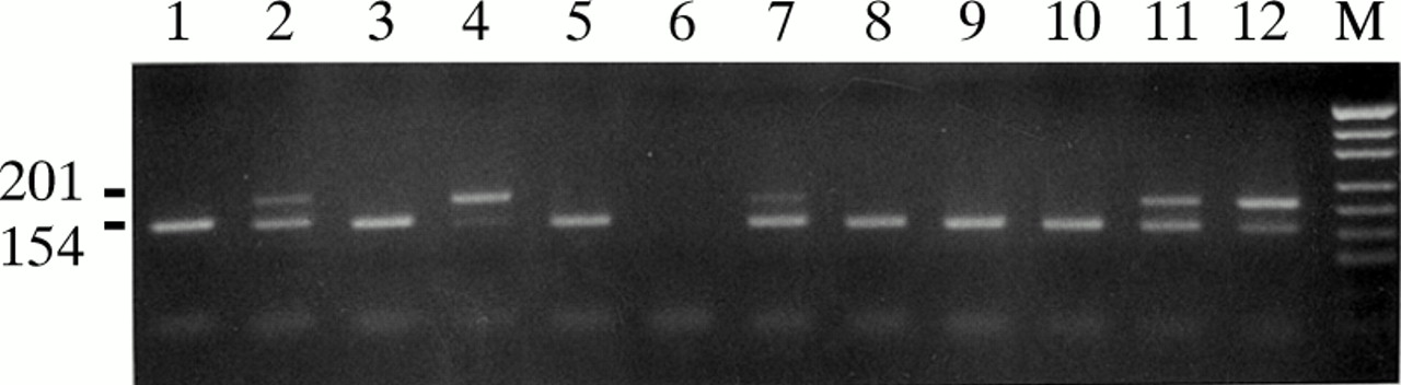
To establish the MSP at the RB1 promoter, 20 tumour DNA samples, which had previously been characterised in detail,1 were used. Characterisation included genotyping with intragenic markers, DNA sequencing, and analysis of the methylation status by Southern blot hybridisation of genomic DNA digested with methylation sensitive restriction enzymeBssHII.3 All tumours showed loss of heterozygosity (LOH) at the intragenic polymorphic loci RBi2 and RB1.20, and an inactivating mutation was identified in the remaining allele. In eight tumours a methylated allele was identified by Southern blot hybridisation. In each of these samples, a 201 bp PCR product was clearly visible when tested in the MSP assay (fig 2, lanes 2, 4, 7, 11, and 12). As expected, DNA from tumour and blood cells not containing a methylated RB1 allele produced a 154 bp PCR product only (fig 2, lanes 1, 3, 5, 8, 9, and 10). No PCR product was obtained from unmodified genomic DNA under the conditions used here (data not shown). Trace amounts of the 154 bp PCR product were also detectable in tumours with LOH at intragenic loci. This finding might be the result of an unhomogeneous cell population consisting of cells with varying degrees of hypermethylation and non-tumour cells. Therefore, the presence of a 154 bp PCR product in the RB1-MSP assay does not necessarily indicate the presence of an unmethylated RB1allele in the tumour. A 201 bp PCR product specific for the methylated allele, however, was detected only in tumours that also showed a methylated allele by Southern blot hybridisation.
MSP analysis of hypermethylated (lanes 2, 4, 7, 11, and 12) and unmethylated (lanes 1, 3, 5, 8, 9, and 10) retinoblastomas; PCR failure (lane 6); marker(M) = MspI digested pUC19 DNA. The positions of the 201 bp PCR product representing the methylated allele and the 154 bp PCR product representing the unmethylated allele are indicated; 5 μg genomic DNA was bisulphite treated as previously described.9 ,11 Amplifications were performed in a reaction volume of 25 μl containing 2 μl of bisulphite treated DNA (Perkin Elmer, PE9600). Conditions: 10 mmol/l Tris-HCl, pH 8.3, 50 mmol/l KCl, 1.5 mmol/l MgCl2, 0.2 μmol/l of each dNTP, 0.25 μmol/l of primer RBcom, 0.3 μmol/l of primer RBmet, 0.05 μmol/l of primer RBunmet, and 0.5 units AmpliTaq (Perkin Elmer). Cycling conditions: 94°C for five minutes for one cycle; 95°C for 15 seconds, 64°C for 15 seconds, and 72°C for 30 seconds for 35 cycles, followed by 72°C for five minutes. PCR products were separated on 3% agarose gels.
To show that the RB1-MSP is applicable in routine diagnosis, a second set of 20 randomly selected tumour DNA samples with unknown methylation status was tested by theRB1-MSP. All tumours had been investigated for LOH at RBi2 and RB1.20. In two of these samples, a methylatedRB1 promoter region was identified by the MSP assay and verified by Southern blot analysis. Although this is a small number of tumours, the finding of methylatedRB1 alleles in 10% of unilateral sporadic retinoblastomas is in agreement with previous estimates on the frequency of hypermethylated RB1alleles.4 An additional 154 bp PCR product representing an unmethylated allele was also obtained in these two tumours, which was to be expected as they did not show LOH at the intragenic polymorphic loci RBi2 and RB1.20.
In summary, analysis of 40 samples has shown that our MSP reliably identifies tumours with hypermethylated RB1alleles as detected by Southern hybridisation. Compared to Southern blot analysis, however, MSP is faster, can be performed with small amounts of genomic DNA, and does not need radioactive components. Thus, MSP facilitates identification of RB1 gene hypermethylation in RB and other tumours.12
Acknowledgments
Part of this work was supported by the Deutsche Krebshilfe. We thank Martina Klutz and Christina Lich for technical assistance.