Article Text

Statistics from Altmetric.com
Fetal growth is a complex and dynamic process regulated by a large number of interactive factors of fetal, maternal, and placental origin. As a result, any abnormality of fetal growth has a complex multifactorial pathogenesis. It has been estimated that about 50% of intrauterine fetal growth is determined by fetal genes.1Maternal disease, her nutritional intake and behaviours, such as smoking, also influence fetal growth. The placenta develops to its full size during the second trimester, to facilitate the fetal growth acceleration after 20 weeks of gestation. An abnormal pattern of placental growth earlier in gestation may result in abnormal fetal growth in the late second or third trimesters.
Key Messages
-
IUGR and SGA occur in about 10% of pregnancies and both are associated with an increased risk for perinatal morbidity and mortality.
-
Environmental and genetic factors are known to be associated with IUGR, although the aetiology of most IUGR remains unexplained.
-
In over 20% of pregnancies with idiopathic IUGR, chromosomal mosaicism confined to extra embryonic tissues (CPM) has been observed. The type of CPM (I–III), the particular chromosomal involvement and the origin of trisomy (mitotic/meiotic) in the aneuploid clone correlate with specific pregnancy outcomes. When meiotic CPM involving trisomy is detected in the placenta, the fetus/neonate has a high risk of having uniparental disomy for that chromosomal pair which is trisomic in the placenta.
-
It is important to study placentas from pregnancies with idiopathic IUGR for the presence of CPM using molecular cytogenetic methods.
-
Detection of CPM not only explains the pathogenesis of fetal IUGR but also provides important information for postnatal follow up.
A recently described genetic condition, confined placental mosaicism (CPM), has been shown to cause clinically significant intrauterine growth restriction (IUGR) or even intrauterine fetal death. CPM is the most common form of constitutional chromosomal mosaicism which is defined as at least two cell lines with different chromosomal complements in a fetoplacental unit derived from a single zygote. In CPM only the placenta is affected unlike in generalised chromosomal mosaicism where both the fetus and the placenta are involved. Since the first report associating CPM with idiopathic IUGR in 1983, our understanding of its prevalence and origin, as well as its specific effect on fetal growth, has increased exponentially.2
Intrauterine growth restriction (IUGR)
Abnormal fetal growth represents a major cause of perinatal morbidity and mortality. IUGR is defined as a pathological process that affects normal fetal growth and results in an infant whose growth is less than its genetic potential. In contrast, the small for gestational age (SGA) infant is one whose birthweight is less than a specific cutoff point, based on average weight for a specific gestational age, usually between the 10th and 5th percentile.3-5 It is obvious that for prognostic, counselling, and management purposes it is essential to discriminate between an infant for whom intrauterine growth has been restricted and that for whom being small is normal. Given the existing difficulty in accurate differentiation between IUGR and SGA, it is important to identify the specific causes of reduced intrauterine fetal growth to understand the long term implications of the newborn’s condition.6 7 For the purposes of this article the terms IUGR and SGA follow those in cited references; a number of SGA infants are included in some IUGR studies.
SGA or IUGR neonates have comparatively high levels of perinatal morbidity and mortality.6 7 Recent epidemiological studies suggest a link between reduced fetal growth and the subsequent development of diseases such as diabetes, coronary heart disease, and hypertension.8 9 Fetal adaptation to adverse placental function, of which low birthweight is the most obvious marker, may “imprint” an individual in such a way that internal organs become permanently defective in their structure and function in adult life.
In spite of high quality prenatal care and postnatal morphological examination of the placenta, the cause of abnormal fetal growth in most infants with IUGR remains undetermined. Various environmental and genetic factors, however, have been associated with IUGR. These can be divided into maternal—alcohol, smoking, illegal drugs, malnutrition, essential hypertension, diabetes—fetal—infection, chromosomal abnormalities, multifetal pregnancy, specific genetic disorders—and placental—abruption, abnormal implantation, confined placental mosaicism—risk factors.3 10 To understand better the dynamics of abnormal fetal growth, it is important to determine whether there are identifiable subgroups, such as confined placental mosaicism, within idiopathic IUGR, and whether a specific long term prognosis can be assigned to them.
Developmental aspects of confined placental mosaicism (CPM)
One of the prevailing beliefs dominating the interpretation of the pathogenesis of abnormal intrauterine development has been based on the assumption of a genetic identity between the fetus and its placenta. However, in around 1–2% of viable pregnancies studied by chorionic villus sampling at 10–12 weeks of gestation, a cytogenetic abnormality, most often trisomy, is confined to the placenta and is absent in the fetus.11 12 This genetic diversity in the conceptus is known as CPM. Pregnancies with CPM and a non-mosaic diploid fetus show normal diploid cytogenetic results in amniotic fluid cell cultures in the second trimester. Prenatally diagnosed CPM usually persists throughout gestation and can be demonstrated in the term placenta.13
Based on the origin of the trisomic chromosome in the placenta, CPM can be designated as either mitotic or meiotic (fig 1). In mitotic CPM the trisomic cell line arises from postzygotic mitotic duplication of one chromosome in the progenitors of a placental cell lineage (either trophoblast or extra embryonic mesenchyme) in the developing normal diploid conceptus. In meiotic CPM a trisomic zygote is rescued at the blastocyst developmental stage by loss of the extra chromosome during mitotic cell division in the embryonic progenitor cells, while the progenitors of the placenta remain trisomic.14 Meiotic CPM may be associated with uniparental disomy, which is defined as the derivation of a pair of homologous chromosomes from one parent. In theory, one third of the trisomic zygote rescues are expected to result in fetal uniparental disomy for the chromosomal pair which is trisomic in the placenta (fig 2). In association with CPM, uniparental disomy may also negatively affect intrauterine growth of the fetus.15 The effect of uniparental disomy on prenatal fetal development may be related to either the presence of specific imprinted genes or homozygous gene mutations. Maternal upd2, maternal upd7, maternal upd9, maternal upd15 and maternal upd16 in pregnancies with CPM 2, 7, 9, 15 and 16, respectively, have been reported and related to trisomy zygote rescue.16-20

Mechanisms of origin of mitotic and meiotic CPM. (A) Mitotic CPM arises from a diploid zygote when a postzygotic error occurs in one of the placental cell lineages (trophoblast or mesenchymal stroma). Usually, the placentas with mitotic CPM will have localised trisomic regions and low levels of mosaicism. (B) Meiotic CPM is a result of a trisomic zygote rescue. Fetal karyotype is diploid due to the loss of trisomic chromosome from embryonic progenitors during early embryonic development. At term, placentas with meiotic CPM have high levels of mosaicism or even 100% anueploidy.

Principle of origin of fetal uniparental disomy during trisomic zygote rescue. Note that, in theory, a third of all cases of meiotic CPM develop this.
Categorised according to the specific placental cell lineage exhibiting the abnormal cell line, three types of CPM exist. Placental mosaicism can be confined either to trophoblast (type I), chorionic stroma (type II), or both cell lineages (type III).11 Types I and II are mostly mitotic in origin and type III is mainly meiotic.
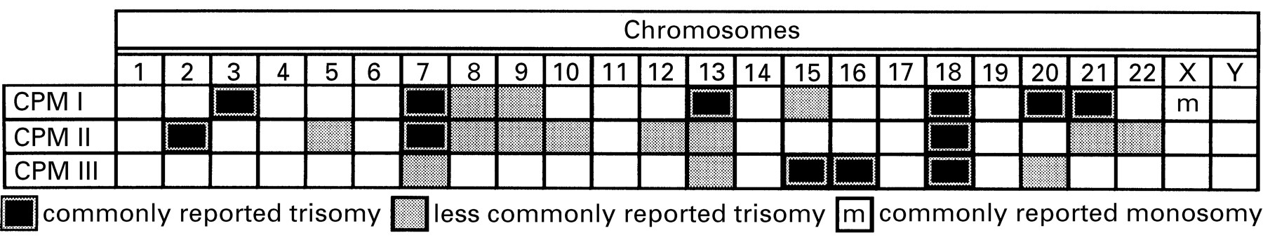
Data from two published collaborative studies show that chromosomes are not randomly involved in the different types of CPM (fig3).12 21 In type I CPM certain chromosomal trisomies, such as 3, 7, 13, 18, 20 and 21, occur more frequently, while 8, 9, and 15 are less common and others are absent. Monosomy in complete or mosaic form is the most frequent finding for the X chromosome. Type II CPM involving the chorionic villus stroma is less common than type I. Frequently seen are trisomies for chromosomes 2, 7, and 18 while trisomies 5, 8, 9, 10 12, 13, 21 and 22 are more rarely associated with this type of CPM. Type III CPM represents trisomic zygote rescue. It has been described many times for trisomies 15, 16, 18, rarely for chromosomes 7, 13, 20 and 22, and not at all for others. In some of the pregnancies with type III CPM, abnormal serum profile (alpha fetoprotein, human chorionic gonadotrophin, uE) in the second trimester has been observed.22

Reported chromosomal involvement for CPM, types I, II, and III, as detected by direct preparation (trophoblast) and long term culture (chorionic stroma) from prenatal chorionic villus sampling. Data were compiled from two major collaborative studies.12 21 Black squares indicate trisomies that have been found in ⩾ 10% of the total of analysed cases of each individual type of CPM. Grey squares indicate trisomies that have been found in ⩾3% but ⩽ 10% of the total of analysed cases of each individual type of CPM.
IUGR and CPM
To what extent does the genetically abnormal placenta influence intrauterine fetal growth and pregnancy outcome? There are two types of pregnancy outcome studies describing either detection of CPM at chorionic villus sampling (10 to 12 weeks of gestation) or in the term placenta of pregnancies associated with SGA and IUGR. Both the timing of placental analysis (CVS vs term placenta) and its completeness (both trophoblast and chorionic stroma analysed) should be considered before correlating CPM with pregnancy outcome.
An increase in pregnancy complications as well as completely normal pregnancy outcomes have been documented in clinical follow up studies of CPM diagnosed at CVS.23 24 The clinical significance of CPM detection is determined by the type of CPM and specific chromosome involvement. Type I CPM is reported to result in spontaneous abortion, IUGR, intrauterine death or perinatal morbidity in 22% of affected pregnancies.23 Type II CPM is mostly found in pregnancies with a normal outcome and rarely with fetal IUGR or intrauterine fetal death.23 In type III CPM, intrauterine death or IUGR are common and most reported cases with intrauterine fetal death are associated with CPM 16.16 25
An abnormal outcome of pregnancy outcome with CPM diagnosed at chorionic villus sampling is usually determined by persistence of high levels of the placental aneuploidy (mostly trisomy) throughout gestation.13 The analysis of term placentas from pregnancies with prenatally diagnosed CPM revealed a 35% prevalence of IUGR when high levels of aneuploid clone were present, while low levels or absence of mosaicism at term correlated with a normal fetal weight.13 26
The prevalence of CPM detected at term in IUGR pregnancies without prenatal CVS diagnosis is reported to be between 8% and 60%.2 25 27 28 The studies including non-idiopathic IUGR pregnancies, such as those with history of heavy maternal smoking, usually detect a low prevalence of CPM.25 On the other hand, when placentas from idiopathic IUGR pregnancies with birthweights below or equal to the 5th percentile are selected, CPM has been detected in 20% or more.2 27 28 Therefore, for greater efficiency of CPM detection in IUGR pregnancies, only placentas from idiopathic IUGR pregnancies without obvious maternal, fetal, and placental causes should be included. Additionally, both placental cell lineages, trophoblast as well as chorionic stroma, should be analysed before excluding a diagnosis of CPM.
The exact mechanism by which abnormal cells in the placenta affect fetal growth and even lead to fetal intrauterine death remains unknown. Although pregnancies with fetal uniparental disomy are often complicated by IUGR, abnormal fetal growth is probably caused by a highly trisomic placenta rather than just uniparental disomy in the fetus. For example, severe IUGR has been reported for placentas with high level CPM 16 in association with both biparental disomy (16) and uniparental disomy (16) fetuses. The same study reported a low level of CPM 16 in a placenta with a normal birthweight infant with upd16.16 As CPM and uniparental disomy often occur in association with one another, the effects of each are difficult to discern when only small numbers of affected pregnancies are reported.
Detection of CPM
To make an accurate diagnosis of CPM all three embryonic cell lineages (trophoectoderm, extra embryonic mesenchyme, embryo proper) should be cytogenetically evaluated. The trophoectoderm lineage is represented by the trophoblast (cytotrophoblast or syncytiotrophoblast). Fibroblasts of chorionic stroma and chorionic plate represent extra embryonic mesenchyme lineage. All fetal cells and the amniotic epithelium are derived from the embryonic progenitors.11
For postnatal investigation of placentas from pregnancies complicated by idiopathic IUGR and fetal death, the most effective approach currently available is comparative genomic hybridisation (CGH). CGH is a technique which offers a molecular approach to cytogenetic analysis and allows the entire genome to be screened for chromosomal imbalances in a single experiment, thus avoiding tissue culture artifacts and culture failures.29 CGH involves co-hybridisation of differentially fluorochrome labelled placental DNA (such as FICT, green) and normal control DNA (TRITC, red) to normal metaphase chromosome preparations. The ratio of the two fluorochromes (green to red) is calculated for each chromosome to determine if there is a chromosomal abnormality in the placenta (trisomy).30 It should be noted that CGH cannot detect balanced chromosomal structural rearrangements and polyploidies and is not reliable for detection of low level mosaicism. However, these limitations are not essential as only high levels of mosaicism seem to be clinically relevant for IUGR. Using CGH accurate data on each placental lineage, IUGR pregnancies of unexplained aetiology can be easily obtained.
When CPM is diagnosed after delivery of an IUGR neonate, DNA analysis of parents, infant, and placenta can determine the origin of placental trisomy and fetal disomy. While a mitotic origin excludes the possibility of fetal uniparental disomy, a meiotic origin of placental trisomy indicates high risk of fetal uniparental disomy for the involved chromosome. It is important for both obstetricians and pediatricians to request appropriate genetic diagnostic tests for neonates and stillborns with idiopathic IUGR to provide the crucial information required for management of IUGR infants and genetic counselling in case of intrauterine death.
Conclusion
The incidence and the phenotypic effects of CPM in human pregnancies with idiopathic IUGR has been underestimated for a long time. To date, both the research and clinical findings show that idiopathic IUGR is probably associated with type III CPM. As the common chromosomal defect in type III CPM is trisomy of meiotic origin, the fetus/newborn carries a risk of uniparental disomy for that chromosomal pair which is trisomic in the placenta. The possibility of clinical detection of uniparental disomy and evaluation of its effect on the postnatal development of the IUGR neonate represents the greatest advantage of saving the placental sample at the time of delivery of a neonate with idiopathic IUGR. Complete placental studies in pregnancies with idiopathic IUGR can unveil the contribution of CPM to fetal growth restriction and teach us about the role of specific chromosomal mosaicism in the regulation of placental and fetal growth.
Acknowledgments
We thank I J Barrett and B L Lomax in the Research Molecular Cytogenetics Laboratory for their technical and editorial assistance. The financial assistance of the March of Dimes Research Foundation (FY96-1034) is gratefully acknowledged.