Article Text

Abstract
In total, 200 families were reviewed with directly transmitted, cytogenetically visible unbalanced chromosome abnormalities (UBCAs) or euchromatic variants (EVs). Both the 130 UBCA and 70 EV families were divided into three groups depending on the presence or absence of an abnormal phenotype in parents and offspring.
No detectable phenotypic effect was evident in 23/130 (18%) UBCA families ascertained mostly through prenatal diagnosis (group 1). In 30/130 (23%) families, the affected proband had the same UBCA as other phenotypically normal family members (group 2). In the remaining 77/130 (59%) families, UBCAs had consistently mild consequences (group 3).
In the 70 families with established EVs of 8p23.1, 9p12, 9q12, 15q11.2, and 16p11.2, no phenotypic effect was apparent in 38/70 (54%). The same EV was found in affected probands and phenotypically normal family members in 30/70 families (43%) (group 2), and an EV co-segregated with mild phenotypic anomalies in only 2/70 (3%) families (group 3). Recent evidence indicates that EVs involve copy number variation of common paralogous gene and pseudogene sequences that are polymorphic in the normal population and only become visible at the cytogenetic level when copy number is high.
The average size of the deletions and duplications in all three groups of UBCAs was close to 10 Mb, and these UBCAs and EVs form the “Chromosome Anomaly Collection” at http://www.ngrl.org.uk/Wessex/collection. The continuum of severity associated with UBCAs and the variability of the genome at the sub-cytogenetic level make further close collaboration between medical and laboratory staff essential to distinguish clinically silent variation from pathogenic rearrangement.
- CGH, comparative genomic hybridisation
- CNV, copy number variation
- DCR, Down’s syndrom critical region
- EV, euchromatic variants
- HAL, haploid autosomal length
- PWACR, Prader-Willi critical region
- TNDM, transient neonatal diabetes mellitus
- UBCA, unbalanced chromosome abnormalities
- transmitted
- unbalanced
- euchromatic variant
- copy number variation
- web site
Statistics from Altmetric.com
- CGH, comparative genomic hybridisation
- CNV, copy number variation
- DCR, Down’s syndrom critical region
- EV, euchromatic variants
- HAL, haploid autosomal length
- PWACR, Prader-Willi critical region
- TNDM, transient neonatal diabetes mellitus
- UBCA, unbalanced chromosome abnormalities
The resolution of the light microscope means that conventional chromosome analysis is limited to the detection of imbalances greater than 2–4 Mb of DNA. Consequently, unbalanced chromosomal abnormalities (UBCAs) usually involve several megabases of DNA, and the great majority are ascertained because of phenotypic or reproductive effects that bring patients to medical attention. The more severely affected an individual, the more likely they are to be investigated, creating an ascertainment bias that does not reflect the full range of phenotypes that may be associated with imbalance of a particular chromosomal segment. In examining subsequent cases, clinicians will naturally tend to look for features already reported and, at the same time, new and unusual features are more likely to reach publication than the absence of previously reported characteristics. Thus, a publication bias may compound a pre-existing ascertainment bias.
Many structural UBCAs are unique in the literature, and the phenotype associated with a given imbalance may depend on a single individual examined at a particular age. As a result, it can take many years before the phenotype associated with a particular imbalance can be defined. However, directly transmitted chromosomal imbalances, where parents and offspring have the same unbalanced cytogenetic abnormalities, provide the means of assessing the phenotype in one or more individuals at different ages as well as the opportunity of judging whether a chromosomal imbalance is a pathogenic or coincidental finding.
These transmitted imbalances are of two contrasting kinds. Firstly, there are the classic UBCAs, in which the copy number of multiple genes is either reduced or increased by one copy as in a deletion or duplication. An increasing number of exceptions to the rule that UBCAs result in significant phenotypic consequences have been reported in families ascertained for “incidental” reasons such as prenatal diagnosis because of maternal age. Secondly, there are the “euchromatic variants” (EVs), which usually resemble duplications. In an increasing number of instances, these reflect copy number variation of segments containing genes and pseudogenes, which are polymorphic in the normal population and only reach the cytogenetically detectable level when multiple copies are present. These EVs segregate in most families without apparent phenotypic consequences. Here, 130 families with transmitted UBCAs are reviewed,1–106 together with a further 70 families107–143 segregating the five established euchromatic variants of 8p23.1,108 9p12,130 9q12 (9qh),113 15q11.2,144 and 16p11.2.128
The 200 families with UBCAs or EVs have been reviewed with respect to the type of rearrangement, size of imbalance, ascertainment, mode of transmission,and the presence or absence of phenotypic effects. Many more cytogenetic and subcytogenetic UBCAs and EVs are being identified now that higher resolution techniques are being used for routine constitutional analysis including high resolution molecular cytogenetics145–147 and array comparative genomic hybridisation (CGH).148,149 Cytogenetically detectable anomalies with little or no phenotypic effect have previously been reviewed only in book form,150,151 and the data from this review have been placed on a web site as the “Chromosome Anomaly Collection” (http://www.ngrl.co.uk/Wessex/collection.html).
METHODS
The contents of this review have been accumulated over time and are thought to contain the majority of documented transmitted UBCAs and EVs. However, there is no systematic way of searching the literature for transmitted anomalies, thus no claim can made that this review is comprehensive.
Criteria for inclusion
Families were selected on the basis of the direct vertical transmission of euploid autosomal UBCAs, or EVs from parent to child. As a result, aneuploid karyotypes were excluded, with the exception of a number of unbalanced tertiary monosomies resulting in transmitted karyotypes with 45 chromosomes. Satellited autosomes have not been included but are reviewed elsewhere.152 Supernumerary marker and ring chromosomes were excluded because of the confounding effects of a high degree of mosaicism on the phenotype.153–155 Transmitted imbalances of the sex chromosomes were also excluded because of the confounding effects of X inactivation in females.
Groups
The UBCA and EV families were divided into three major groups depending on the presence or absence of a detectable phenotypic effect in offspring, parents or both (table 1). Group 1: families in which transmitted UBCAs or EVs had no apparent phenotypic consequences in probands, parents and other family members; group 2: families in which the same UBCA or EV was found in affected probands as well as phenotypically normal parents and other family members; and group 3: families in which the same UBCA or EV was found in affected probands as well as affected parents and other family members.
Phenotypic normality
Individuals were considered phenotypically affected when any type of phenotypic anomaly was mentioned even if the aetiological role of the chromosome abnormality in the same individual is questionable. It is acknowledged that individuals in a given family may not have necessarily been examined by clinical genetic staff, but patients were presumed normal unless otherwise stated.
Size of imbalances
Wherever stated, estimates of the size of the imbalances derived by the authors of the relevant papers were used. Elsewhere, the size of each imbalance was estimated by measuring the proportion of the normal chromosome represented by the deleted or duplicated material on high resolution standardised idiograms and multiplying by the % haploid autosomal length (HAL) of the chromosome concerned.156 The % HAL was converted to Mb by multiplying by the 2840 Mb estimated length of the human genome.157
RESULTS
The review covers 200 families in which 130 had transmitted UBCAs and 70 had transmitted EVs.
Transmitted unbalanced chromosome abnormalities
The location and extent of the UBCAs is illustrated in fig 1, and details of the 130 UBCA families in groups 1, 2, and 3 are listed in Appendices 1, 2, and 3. Table 1 provides a summary of the ascertainment and the sex of the transmitting parents in each group and table 2 summarises the size of the imbalances.
The 130 families contained 374 UBCA carrying individuals with 111 different transmitted autosomal rearrangements involving 20 of the 22 autosomes, the exceptions being chromosomes 12 and 17. Chromosomes 5, 8, and 18 were the most frequently involved. Independent confirmation by FISH or molecular methods had been obtained in more than half (87/130 or 67%) of the families.
Over half these families (77/130 or 59%) fell into group 3, in which a degree of phenotypic expression is found in both children and parents. Approximately a quarter fell into group 2 (30/130 or 23%), in which an affected proband has the same UBCA as an unaffected parent, and the remaining one fifth made up group 1 (23/130 or 18%), in which neither children nor parents are affected. Many of these imbalances were unique to the family concerned.
Group 1: Phenotypically unaffected parents with the same unbalanced chromosome abnormality as their unaffected children
This group contained 23 families in which an unbalanced rearrangement had been directly transmitted from parent to child without phenotypic effect in 66 carriers. For completeness, four chromosomally unbalanced but phenotypically normal individuals were included from families in which direct transmission from an unbalanced parent had not been observed,158–161 making a total of 27 families. The majority (20/27; 74%) of these families was ascertained at prenatal diagnosis because of maternal age (12/20). Of the remaining seven (17%), three were ascertained for miscarriages,2,9,158 three because of the phenotype of a sibling20,160,161 or daughter,159 and one for infertility.14
Of the 27 families, 14 had deletions, with an average size of 8.2 Mb (range 4.2–16.0 Mb) (table 2), and of these, 12 consisted mainly of G dark bands with or without some G light flanking material. Seven families had transmitted interstitial duplications with an average size of 13.6 Mb (range 3.4 Mb to 31.3 Mb), of which only the duplications of 8p2215 and 13q14-q2117 were largely G dark bands. There were six families with unbalanced rearrangements, three of which had been transmitted from a parent with the same imbalance19–21 and three from a parent with a balanced form of the same rearrangement.159–161
In the 23 families in which the UBCA had been directly transmitted from a parent to child, table 1 shows that the transmission was maternal in 15 families (71%), paternal in in five, (22%), and from both parents in three (13%).
Group 2: Unaffected parents with the same autosomal imbalance as their affected children
This group contains 30 families with 78 carriers (Appendix 2). The majority (25/30; 83%) were ascertained because of phenotypic abnormality (PA) in the proband. Of the remaining 5 (17%), two were ascertained because of the phenotype of a sibling proband,30 one because of infertility,42 one because of leukaemia27 and one as a result of prenatal diagnosis following an abnormal ultrasound scan.31
Seven families had transmitted deletions with an average size of 7.5 Mb (range 3.6–10.0 Mb) (table 2) of which three largely involved the G dark bands 5p14 and 11q14.3. Nineteen families had transmitted duplications with an average size of 6.1 Mb (range 2.0–16.3 Mb) of which the duplications of 4q3230 and 8p23.232 were mainly G dark. Three families had transmitted unbalanced translocations.
Table 1 shows that exclusively maternal transmission was seen in 19/30 families (63%) of families, paternal in 9/30 (30%), and from both in 2/30 (7%).
Group 3: Affected parents with the same autosomal imbalance as their affected children
This group contains 230 carriers from 77 families (Appendix 3). Of 77 families, 71 (92%) were referred for phenotypic abnormalities in the proband, which were, in most cases, reflected to a lesser or greater extent in other carriers from the same family.
Four of the 77 families (5%) were ascertained through prenatal diagnosis; two of these because of maternal age,58,91 one because of abnormal ultrasound,67 and one because of a previous son with mental retardation.65 A single family was investigated because of miscarriages106 and a single family because of Prader-Willi syndrome in the proband.33
Thirty-eight families out of 77 (49%) had deletions with an average size of 10.9 Mb (range 2.0–30.8 Mb). Twenty-seven families (35%) had transmitted duplications with an average size of 11.0 Mb (range 4.0–26.1). The remaining 12 (16%) had transmitted unbalanced translocations of which 4 were insertional.
Table 1 shows that exclusively maternal transmission was seen in 58/77 families (75%) of families, paternal in 9/30 (16%) and transmission from carriers of both sexes in 7/77 (9%).
Group 1 and 2 UBCAs, especially those overlapping with Group 3
Brief summaries are provided here of all group 1 and 2 UBCA families. Group 3 families are included wherever group 3 UBCAs overlapped with group 1 and/or group 2 UBCAs (fig 1).
der(1)(p32-pter)
One unconfirmed monosomy of 1p32 to pter was ascertained at prenatal diagnosis and also apparently present in the father.19 This UBCA, reported in abstract, is impossible to reconcile with a normal phenotype, as even small imbalances of distal 1p are associated with a recognisable chromosomal syndrome.162
dup(1)(p21-p31)
This large group 1 duplication was ascertained at prenatal diagnosis for maternal age. The duplication was found in the phenotypically normal mother, and the outcome of pregnancy was normal at term.13
dup(1)(q11-q22)
This group 2 family was ascertained in a phenotypically normal boy of 9 with lymphadenopathy.27 A constitutional duplication of proximal 1q was found in this boy, his phenotypically normal mother and his elder sister, neither of whom had lymphoma or leukaemia.
dup(1)(q42.11-q42.12)
This group 2 family was ascertained in a boy who fed poorly and was in the 10th centile for growth.28 The duplication had arisen de novo in the phenotypically normal mother and, by the age of 3 years, the boy’s stature was in the 25th centile when correlated with the height of his parents.
del(2)(p12-p12)
Two group 1 families with deletions of 6.1 Mb and 6.7 Mb within G dark 2p12 were both ascertained at prenatal diagnosis.1 At least 13 loci including a cluster of six pancreatic islet regenerating genes were deleted. The pregnancies had normal outcome at birth and there were no other apparent phenotypic consequences in six other deletion carriers. It was proposed that segmental haplosufficiency may be associated with low gene density, especially where genes within a cluster on the normal homologue may compensate for each other, or genes of related function are present on other chromosomes.25 An overlapping 7.5 Mb group 3 deletion extended into the gene rich part of 2p11.2 and was found in a girl with speech delay and in her mother, who has expressive language difficulties (patients 25147 and 31). Both had mild dysmorphic features.
del(2)(q13-q14.1)
A group 1 family was ascertained because a woman of 38 years had three early miscarriages. The deletion spanned 7 cM from YAC 791f4 to YAC 676d2. The consultand and her phenotypically normal mother had the same deletion, but the mother had no history of miscarriage.2
del(3)(p25-pter)
A terminal group 1 deletion with a 3p25.3 breakpoint was ascertained at prenatal diagnosis in a fetus and phenotypically normal mother.3 In contrast, in a group 3 family, an affected boy and his less severely affected mother had features consistent with 3p-syndrome.46 It was suggested that the 3p25.3 breakpoint was distal to the genes responsible for 3p-syndrome.3 However, this could also be an example of non-penetrance of a chromosomal deletion, as haploinsufficiency of the CALL gene is thought to give rise to mental impairment and this gene should lie inside the deletion at 3p26.1.163
dup(3)(q25-q26)
A group 2 family contained two sisters with congenital heart disease, mild developmental delay, dysmorphic, features and a dup(3)(q25q25).29 The same duplication was present in the normal father, grandmother, and greatgrandmother. The authors suggested a paternal imprinting effect, but this region of chromosome 3 is not known to be imprinted. A group 3 family with a larger overlapping dup(3)(q25.3q26.2) was independently ascertained once with congenital heart disease and once with microcephaly.78 These families suggest that the phenotype associated with duplication of 3q25 can extend into the normal range or that 3q25 contains a dosage sensitive locus that gives rise to heart disease with variable penetrance.
dup(3)(q28q29)
A group 1 family was ascertained at prenatal diagnosis for maternal age and found in the phenotypically normal father and an older sibling.12 A submicroscopic duplication of 3q29 was ascertained in siblings with moderate mental retardation and dysmorphic features164 but was also present in the phenotypically normal mother and sister.
dup(4)(q31-q32)
A group 3 family with a duplication of 4q31.1-q32.3 was ascertained in a mildly affected child and his mother, who were both developmentally delayed.79 This prompted Maltby et al30 to report a smaller group 2 duplication of 4q32 ascertained because of trisomy 21 in the proband. The duplication carrying sister had sensorineural deafness and the mother had no obvious clinical problems. The authors concluded that there were insufficient consistent findings to suggest a clinical effect, but this family also suggests that overlapping duplications centred on G dark 4q32 have a variable phenotype that can extend into the normal range. Few clinical details of the group 3 family of Van Dyke77 were given.
del(5)(p15-pter) terminal
There were two group 2 deletions of 5p15.3 and 10 group 3 monosomies of this region. The group 2 families had microcephaly, a cat-like cry and developmental delay, but not the severe delay and facial features of cri du chat syndrome associated with deletions of 5p15.2.22 There were four affected children in these group 2 families, but the carrier parent was apparently normal in each case. “Atypical” cri du chat syndrome in parents and children has also been described.51–55 These families suggest a variable phenotype that can extend into the normal range but is more often characterised by speech delay, occasional deafness, and low to normal intelligence.
del(5) (p13-p15) interstitial
There were one group 1 and two group 2 deletions of 5p14 itself as well as four larger overlapping group 3 deletions. The group 1 deletion of almost all 5p14 was ascertained at prenatal diagnosis and found in a total of six normal carriers.4,23 The G dark 5p14.1-5p14.3 group 2 deletion ascertained in a patient with a peroxisomal disorder was thought to be an incidental finding, as this condition had not previously been associated with any case of 5p deletion.10 In a more recent family,23 a non-mosaic deletion contained within 5p14 was found in a proband with microcephaly, seizures, and global developmental delay; the phenotypically normal father had the same deletion in blood, but only 1/500 fibroblasts. Nevertheless, given the eight carriers in the other two 5p14 deletion families and the normal phenotype of the father, it seems likely the proband in this family represents ascertainment bias rather than variable expression of a phenotype associated with this deletion. By contrast, all the four overlapping group 3 deletions extended into adjacent G light 5p13, 5p15 or both. The phenotype varied within and between families from mild21 to variable57,58 and severe in the family of Martinez et al,56 which showed that cri du chat syndrome is compatible with fertility.
dup(5)(q15-q22.1)
A group 2 family with a dup(5)(q15q21) was ascertained at prenatal diagnosis because a cystic hygroma was found in one of two monzygotic twins using ultrasound.31 The authors concluded that the dup(5) could be a coincidental finding in view of the discordant abnormalities in the twins after delivery and the normal phenotype of the father. However, the father had suffered from epilepsy as a child and it is not unknown for cytogenetic abnormalities to have different consequences in monozygotic twins.165 A larger overlapping group 3 duplication also had a variable phenotype with mild dysmorphic features in mother and son but no mental retardation in the mother.80
dup(6)(q23.3-q24.3)
Both the group 2 families were ascertained with transient neonatal diabetes mellitus (TNDM) and have duplications that include the paternally imprinted ZAC locus, which maps to 6q24.2. Imprinting explains the presence of TNDM in carriers with paternal duplications and the absence of TNDM in carriers with maternal duplications. While the proband and father in the family of Temple et al41 were discordant for TNDM, a degree of developmental delay in the father is probably due to this inserted duplication extending beyond band 6q24. An exceptionally mild phenotype was associated with an overlapping de novo 4–5 Mb deletion of 6q23.3-q24.2 that was of paternal origin.166
del(8)(p23.1/2-pter)
A group 1 family with a del(8)(p23.1-pter) deletion was ascertained at prenatal diagnosis in a fetus and phenotypically normal father.5 The deletion breakpoint was believed to be more distal than the de novo deletions associated with developmental delay and heart defects. However, a group 3 family with an 8p23.1-pter deletion was ascertained in a boy of 7 years with mental slowness, behavioural problems, and seizures.59 His sister and father had minimal phenotypic abnormalities with borderline to normal intelligence. A de novo terminal deletion of 8p23.1-pter was ascertained in a girl with initial motor and language delays but average cognitive development and intellectual ability after close monitoring over a period of 5 years.167 These examples indicate that distal 8p deletions are associated with a mild phenotype that can extend into the normal range.
del(8)(q24.13q24.22)
This group 1 family was ascertained because of a positive triple screen test.6 The phenotypically normal mother had the same deletion and a history of miscarriage and fetal loss. The pregnancy with the deletion resulted in a 26 week phenotypically normal stillbirth with significant placental pathology.
dup(8)(p23.1p23.3)
A group 1 family was ascertained for oligoasthenospermia, which was regarded as incidental in view of the normal fertility of a male carrier relative.14
dup(8)(p23.1p23.2): the abnormalities in the probands from three independent group 2 families with 2.5 Mb duplications of G-dark 8p23.2 were inconsistent and not present in any of the carrier parents.32 The authors concluded that duplication of G-dark 8p23.2 could probably be described as a benign cytogenetic variant.
dup(8)(p23.1p23.1)
There were 3 group 2 families and 3 group 3 families with cytogenetic duplications of 8p23.1.33,84 The abnormalities in the probands of the 3 group 2 families were inconsistent with each other and the same duplication was present in one of the parents in each family with no reported phenotypic abnormalities. In the 3 group 3 families, the first was ascertained with developmental delay while the carrier mother had short stature and abnormal feet.33 The second had Prader-Willi syndrome as well as an 8p23.1 duplication while the duplication carrier father had only atrial fibrillation.33 The third group 3 family was a developmentally normal girl of 16 with a severe congenital heart defect.34 The authors proposed that her duplication interrupted the GATA-Binding Protein gene (GATA4), which maps to 8p23.1 and is known to give rise to heart defects when deleted. Her father had an isolated right aortic arch and his milder heart defect was attributed to mosaicism for the duplication. However, these cytogenetic duplications bear an uncanny resemblance to the EVs of 8p23.1 (see below), which have been shown to result from copy number expansion of a discrete domain within band 8p23.1 that does not contain the GATA4 locus.108,109 Thus, apparent duplications of 8p23.1 have been associated with a wide variety of presentations but, as the content of many of these imbalances has not yet been determined, ascertainment bias may account for some of these observations and further analysis could distinguish genuine cytogenetic duplications from euchromatic variants of 8p23.1.
dup(8)(p21.3-p23.1), (p22-p23.1) and (p21.3-p22 or p22-p23.1)
Developmental or speech delay has been associated with duplications of 8p21.3-p23.1 in 2 group 3 families.86 Family 1 was ascertained with a complex heart defect but the mother and a sibling had the same duplication and no heart defects. Family 2 was ascertained for speech delay in a girl who had an IQ of 71 at age 6 and minor facial anomalies. Her carrier sister also had speech delay as well as a heart defect and mild facial dysmorphism. The normal phenotype in her father was attributed to mosaicism for the duplication, which was present in 6/24 cells. The authors concluded that this duplication is associated with mild to moderate delay without significant or consistent clinical features. A similar phenotype was reported in the group 3 duplications of 8p22-p23.1.85,87
dup(8)(p22-p22)
A group 1 family with a small, “euchromatic expansion” of distal 8p22 was ascertained at prenatal diagnosis, confirmed with CGH and found in the phenotypically normal mother and grandfather.15 Overlapping de novo duplications of 8p22-p23.1 were recently reported using high resolution CGH in six families and thought to have Kabuki make-up syndrome168 but these observations have not been replicated by others.169
del(9)(p12.2p22.1)
A group 1 family was ascertained at prenatal diagnosis for maternal age when this deletion was found in the fetus as well as the phenotypically normal father and grandmother.7
dup(9)(p12-p21.3)
A neonate ascertained with cri-du-chat syndrome had a deletion of chromosome 5 derived from her father who had an unbalanced insertional duplication of 9p12-p21.3.159 The estimated size of the duplication was 21 Mb including approximately 280 genes. The balanced ins(5;9)(p13.3;p12p21) form of this insertion was present in the proband’s grandmother and uncle.
del(10)(q11.2-q21.2)
This deletion was found in the clinically normal 29 year old male partner of a couple referred for recurrent miscarriages.158 A patient with an overlapping de novo deletion had normal physical and psychomotor development until the age of 6 but subsequently developed symptoms of Cockayne syndrome. As the excision repair gene (ERCC6) associated with the autosomal dominant type II Cockayne syndrome has been mapped to band 10q21.1, it seems that deletion of proximal 10q is compatible with a normal phenotype but only if the ERCC6 locus is excluded or non-penetrant.
dup(10)(p13-p14)
This group 1 family was ascertained at prenatal diagnosis in a family with a history of heart disease.16 The duplication was found in the fetus with normal outcome at birth, the phenotypically normal mother and a further child who had Tetralogy of Fallot (TOF). Other family members had TOF without the duplication of 10p13 and the authors concluded this is a duplication without phenotypic consequences.
del(11)(q25-qter)
The der(11)t(11;15) Group 2 family was ascertained for infertility.42 No phenotypic anomalies were reported in either the proband or his father but 61% of spermatocytes in the proband had XY multivalent contact at prophase suggesting a causal connection between the unbalanced translocation in the son despite the evident fertility of his father. Unpublished observations from this laboratory include another group 2 deletion of most of 11q25 ascertained in a boy of 6 with developmental delay (especially speech) but no heart defect. His phenotypically normal father had the same deletion. The larger overlapping group 3 deletion of 11q14.2-qter61 was ascertained in a child of nearly 3 with developmental delay. She also had a VSD but a heart defect was not suspected in the mother. Until more of these deletions have been mapped at the molecular level, it is impossible to say whether the phenotypically normal family members with 11q25 deletions are examples of segmental haplosufficiency or a variable phenotype that extends into the normal range. A second group 2 family in which an unbalanced der(11)t(11;22) translocation is dealt with under del(22q) below.43
del(13)(q14q14), dup(13)(q14.1q21.3) and dup(13)(q13-q14.3)
A group 2 family with a deletion of 13q14 was ascertained with retinoblastoma.26 A larger overlapping group 3 deletion was associated with both retinoblastoma and dysmorphic features in a mother and child.62 As retinoblastoma is recessive at the cellular level, the lack of a ‘second hit’ is likely to explain the absence of retinoblastoma in the mother of the first family.26 In a third family, unbalanced segregation of a balanced maternal ins(20;13)(p12;q13q14.3) insertion resulted in deletion of 13q13-q14.3 and retinoblastoma in the proband.160 However, the proband’s older sister had a duplication of the same segment and was clinically normal as was a younger sister at birth.
del(13)(q21q21) and dup(13)(q14-q21)
A group 1 del(13)(q21q21) was ascertained for recurrent miscarriages in a phenotypically normal family.9 An overlapping group 1 dup(13)(q14-q21) was detected at prenatal diagnosis when an extra 13q14 LIS1 signal was seen in interphase cells and only a partial duplication of chromosome 13 in metaphases.17 The same duplication was present in the mother who was clinically normal apart from hyposomia.
dup(13)(q14-q21) and dup(13)(q13-q14.3)
See del(13) entries above.
dup(14)(q24.3-q31)
In a group 2 family, imprinting might have explained the normal phenotype in the father of a girl who had developmental delay, microcephaly and dysmorphic features at the age of 3½ effects.34 However, grandmaternal transmission could not be established as the father was adopted. In addition, the girl had only a few of the features recorded in previous cases of pure 14q duplication. It is therefore impossible to be certain whether the dup(14) is the cause of the child’s phenotype or an incidental finding in this family.
dup(15)(q11.2q13)
There are at least five group 235–39 and four group 3 families35,93 with transmitted interstitial duplications that include the PWACR. The imprinted nature of this region explains the fact that children with developmental delay and/or autism all had maternal duplications35–39 while the normal parents in three of these five families had duplications of grandpaternal origin.37–39 Both parents and children were affected in the four group 3 families35,93 but two out of three unaffected grandparents again had duplications of grandpaternal origin.35 However, one mother with a paternally transmitted duplication had mild developmental delay and it is therefore possible that the phenotype associated with paternal duplications can extend into the mildly affected range. Bolton et al35 compared the phenotype of 21 individuals from 6 families and found that maternally transmitted dup(15)(q11.2q13) was associated with a variable degree of intellectual impairment and motor coordination problems but only one individual met the criteria for classic autism.
del(16)(q21q21)
Two independent group 1 families were both ascertained at prenatal diagnosis with deletions of G-dark 16q21.10,11 There were two other phenotypically normal carriers in each family. The family of Witt et al11 has previously been contrasted with an adult patient who had a cytogenetically identical deletion of 16q21170 but many of the features of 16q- syndrome.171
dup(16)(q12.1q12.1), (q11.2-q12.1) and (q11.2-q13.1)
Verma et al40 considered a duplication of 16q12.1 in an autistic child of 4½ and his clinically normal mother as an unusual variant. The overlapping duplications of q11.2-q12.1 and q11.2-q13.1 were consistently associated with developmental delay, speech delay, learning difficulties and behavioural problems21,94 while de novo adult cases have been associated with a more severe phenotype.172 In most of these families, the duplicated material is found within the major 16q11.2/16qh block of heterochromatin but these are clearly not analogous to the EVs of 9q12/9qh (see below). It seems that duplications of proximal 16q can be severe but are more often associated with a variable cognitive phenotype that may exceptionally extend into the normal range.
del(18)(cen-pter)
There were a total of 7 families with transmitted deletions of 18p including a single group 1 family with a deletion of 18p11.31-pter12 and 6 group 3 families with deletion breakpoints that ranged from p11.365 to the centromere.70 The group 1 family was ascertained at prenatal diagnosis for a raised serum AFP and had the smallest deletion. The group 3 family of Rigola et al65 was ascertained at prenatal diagnosis because of a previous son with mental retardation. The authors concluded that the phenotype in their 18p11.3-pter deletion family was subtle as the mother had only mild mental retardation and minor congenital malformations. In another group 3 family,66 both the child and mother with del(18)(p11.21-pter) had short stature, mental retardation and ocular anomalies. By contrast, the group 3 del(18)(p11.2-pter) of Tonk and Krishna67 was ascertained because of abnormal routine ultrasound findings. A very dysmorphic fetus with features that included cyclopia was found after spontaneous delivery at 24 weeks gestation while the mother had mild mental retardation and some dysmorphic features but. Concordant phenotypes with many of the features of 18p- syndrome were seen in the other three group 3 families with larger 18p deletions.68–70
dup(18)(cen-pter)
A group 1 family with a duplication of the whole of 18p was ascertained at prenatal diagnosis following a raised serum AFP.18 At 2 years of age, the child’s development was normal and she shared bilateral short fifth fingers with her carrier mother and pre-auricular pits with her father. After reviewing 14 other cases, the authors concluded that duplication of 18p produced little if any phenotypic effect. By contrast, Moog et al95 ascertained a group 3 family with a duplication of the whole of 18p in a child with psychomotor delay, slight craniofacial anomalies and moderate mental retardation. The mother had the same duplication in 80% of cells and had been developmentally delayed. By the age of 26, she had height and head circumference less than the 3rd centile and “borderline” mental impairment. The father was also mentally retarded. The authors concluded that duplication of 18p is not a specific phenotypic entity but may be associated with non-specific anomalies and a variable degree of mental impairment. Thus, duplication of 18p has mild phenotypic consequences that can extend into the normal range.
dup(18)(q11.2q12.2)
This duplication was found in the fetus of a mother of 24 referred for prenatal diagnosis with a family history of Down’s syndrome.161 The mother and her next child had a balanced ins(18)(p11.32;q11.2q12.2) insertion but a third child had the corresponding duplication and was phenotypically normal at three months of age.
del(21)(q11.2-q21.3),(pter-21q21.2), (pter-q21)
A group 1, group 2 and group 3 family were each ascertained as a result of Down’s syndrome in the proband. In each family, tertiary monsomic forms of unbalanced translocations were found in two or more other family members. In the group 1 family,20 there were no reported phenotypic anomalies in four family members. However, it is possible that this fusion of 6p and 21q involved no actual loss of coding material especially as de novo loss of subtelomeric 6p has been associated with mental retardation, dysmorphic features and a heart defect.163 In the group 2 family, an unbalanced 19;21 translocation with deletion of pter-q21.1 and a possible deletion of 19p was ascertained in a child because of Down’s syndrome in a sibling proband.44 The child had only behavioural difficulties and the carrier mother was of average intelligence. In the group 3 family, four family members had a complex unbalanced 21;22 translocation and effective monosomy for 21q21.2-pter.104 This family had a consistently mild phenotype with developmental delay, learning disabilities and poor social adjustment. The only group 3 deletion of the 21q11.2-q21.3 region75 was ascertained in a child with dislocation of the hips at 11 months of age. By the age of 5 he had motor and language delay and the mother had mild mental retardation. The authors concluded that psychomotor retardation is the only consistent feature of proximal 21q deletion with a variable degree of expression of other minor anomalies. Roland et al75 also pointed out that more severe de novo cases have been reported as well as a de novo case with normal intelligence but poor motor skills.173 A duplication of proximal 21q with normal phenotype has also been reported.174
del(22)(q11.21-pter)
In the group 1 family, an unbalanced tertiary monosomic (9;22) translocation was ascertained during prenatal diagnosis and found in three other family members.21 The 9q subtelomere was intact, but a diminished signal from BAC 609C6 indicated a 22q11.21 breakpoint and the loss of some coding material from proximal 22q. In the group 2 family, an unbalanced der(11)t(11;22) tertiary monosomy was ascertained in a dysmorphic boy with a heart defect, his two siblings, and his mother.43 The phenotype could have resulted from the deletions of either 11q25 and proximal 22 or both. As only one of the two siblings had a heart defect and the mother was clinically normal, the authors suggested that the unbalanced karyotype might be a coincidental finding in view of the variability of the phenotype. However, variable expression of heart defects is now well known in transmitted submicroscopic deletions of 22q11.2101,175 and suspected in 11q25 deletions (see del(11)(q25-qter) above). In the group 3 family, an unbalanced der(4)t(4;22) translocation and monosomies of both 4q35.2-qter and proximal 22q were ascertained in a dysmorphic boy with a heart defect.101 The complete and partial Di George syndrome seen in the son and mother was attributed to the proximal 22q deletion, although heart defects have subsequently been described in other unbalanced submicroscopic translocation involving 4q.163
Euchromatic variants
The cytogenetic locations of the five major EVs are illustrated in fig 2, and the details of 70 EV families in Appendices 4, 5, and 6. By contrast with the UBCA families, each of these EVs has been independently ascertained on multiple occasions. Of the 70 families, 38 were group 1 (54%), 30 were group 2 (43%), and only two were group 3 (3%). Table 1 provides a summary of the ascertainment and sex of the transmitting parents in each group. The EVs of 8p23.1, 15q11.2, and 16p11.2 have been described as constitutional cytogenetic amplifications because they involve variable domains that are only detectable at the cytogenetic level when present in multiple copies.109,120,133,177
Group 1 EVs: Phenotypically unaffected parents with the same EV as their unaffected children
This group contains 38 families with 94 carriers involving all five of the most common EVs established to date (Appendix 4). Of the 38 families, 30 were ascertained at prenatal diagnosis (79%),12 of whom had undergone the procedure because of maternal age. Four families were referred for recurrent miscarriages and one for loss of a pregnancy, but it is difficult to reconcile this with phenotypically silent EV unless such variation predisposes to larger imbalances or non-disjunction of the same chromosome; this has not been shown in any of the families listed here to date. Two families were investigated because of trisomy 21 in a relative and the final family was ascertained incidentally during a survey of newborns.113
Table 1 shows that exclusively maternal transmission was seen in 18 of the 38 families (47%) of families, paternal in 17 (45%), and transmission from both in three families (8%).
Group 2 EVs: Unaffected parents with the same EV as their affected children
Appendix 5 contains 84 carriers from 30 families. All 30 were ascertained for dissimilar phenotypic abnormalities in the probands. One family was independently ascertained once in a male of 62 years with myelodysplasia139 and once in a child of 3 years with developmental delay and mild dysmorphic features.128 Six other family members were phenotypically normal and this child was later diagnosed with fragile X syndrome (Thompson, personal communication).
Table 1 shows that exclusively maternal transmission was seen in 13 of the 30 families (43%), paternal in nine (30%) and transmission from both in eight (27%).
Group 3 EVs: affected parents with the same EV as their affected child
There were only two families in this group (Appendix 6). In the first family, an 8p23.1 EV was associated with very mild dysmorphism in a mother and her two daughters; further family members were not available and the association of EV and phenotype remains questionable.142 In the second family,143 short stature cosegregated with a proximal 15q amplification variant that was later shown to involve multiple copies of the proximal 15q pseudogene cassette.176 Apart from short stature, the proband had slight hypotonia and a tendency to hyperphagia but no functional modification of the PWACR could be found. The authors concluded that this EV was probably not related to the child’s phenotype. Transmission was maternal in both families.
Group 1, 2, and 3 EVs especially where these overlap with UBCAs
Brief summaries are provided of the group 1 and 2 EV families with particular attention to those instances where group 1 and 2 EVs overlap with each other or with group 3 EVs (fig 2).
8p23.1v
At least 11 families have been reported with this apparent duplication of 8p23.1 (8 in group 1 EV, 2 in group 2 EV and 1 in group 3 EV). Twenty-five out of the 27 carriers in the first three reports were phenotypically normal.108,110,111 Similar findings were reported in two further families107,129 while only minimal features were found in the single group 3 family.142 Williams et al110 found variation of 8p23.1 in a developmentally delayed boy of 18 months but his delay was said to be “spontaneously resolving” by the age of 2 years (Williams L, personal communication). Hollox et al109 used quantitative multiplex amplifiable probe hybridisation to show that the underlying basis of the duplication in three of these EV families was the increased copy number of a domain of at least 260 kb containing three defensin genes (DEFB4, DEFB103, and DEFB104) and a sperm maturation gene (SPAG11). Semi-quantitative FISH indicated that an olfactory receptor repeat is also involved and a recent contig suggests that this domain is normally within the distal 8p23.1 OR repeat itself (REPD).177 Total copy number of this domain in normal controls varied between 2 and 7, whereas EV carriers had between 9 and 12 copies. Expression of DEFB4 was increased with copy number and, as the defensins encode cationic antimicrobial peptides, it has been suggested that increased copy number could enhance resistance to infection or modify the effects of Pseudomonas aeruginosa in cystic fibrosis.109 Copy number variation of a 1 Mb domain that lies 7 Mb from the telomere (CNP 45) has been detected in normal controls,178 but it is not certain that this coincides with the defensin EV, which is thought to lie at or adjacent to REPD at 7.5 Mb from the telomere. Tsai et al33 and Kennedy et al84 claim that duplications of 8p23.1 are associated with developmental delay and heart disease but have not mapped the extent of their duplications (see UBCA dup(8)(p23.1p23.1) above). Recent evidence submitted for publication179 indicates that duplications and EVs of 8p23.1 resemble each other at the cytogenetic level but can be separated into two distinct groups: (a) genuine 8p23.1 duplications of the interval between the olfactory receptor repeats including the GATA4 gene and associated with developmental delay and heart defects; and (b) EVs that involve increased copy number of the variable defensin domain only and do not have phenotypic conseqences.
9p12v
There are at least eight families with this EV (six group 1 EVs and two group 2 EVs), which resembles a duplication of G dark 9p12 and is negative when C banded. Webb et al112 described the extra material as being of “intermediate density” when G banded, noted how the extent of the extra material can vary when transmitted, and suggested that this EV is a homogeneous staining region. As 9q12 EVs derive from a unit present in multiple copies in both 9p and 9q115,180 (see below), it is likely that the cytogenetic 9p EVs also reflect increased copy number of a variable domain by analogy with the 16p11.2 EVs (see below). It is possible that these coincide with the 9p11 and 9q12 polymorphisms identified by Sebat et al (CNPs 51 and 52).178
9q12v/9qhv
There are at least seven families with this EV, which reflects extra C band negative, G dark material that is found within the major 9q12/qh block of heterochromatin (six group 1 EVs and one group 2 EV). The group 2 EVs had 9q13-q21 breakpoints,132 but resembles the other 9q12/qh EVs at the cytogenetic level. YAC 878e3 hybridises to the extra material in the 9q12/qh EVs, and subclones of this YAC indicate that these EVs derive from a large unit present in multiple copies in both proximal 9p and juxtaheterochromatic 9q13.115,180 A shared identity between subclones and expressed sequence tags suggests that this variation includes coding sequences.180 Sequences of this type may also underlie the unconfirmed claim that a separate type of 9q12v chromosome exists with material derived from 9q13-q21.151
The established 9q12 EVs are clearly not analogous to the extra euchromatic material found within the major 16p11.2/qh block of heterochromatin, which has so far always been a genuine duplication of proximal 16q (see UBCA dup(16) above).
15q11.2v
At least 32 families have been reported with extra material within proximal 15q (10 group 1 EVs, 21 group 2 EVs, and a single group 3 EV family). These EVs resemble duplications or triplications and can be misinterpreted as a duplication of 15q11.2-q13 or even a deletion of the homologous 15. The underlying basis of this EV is variation in the copy number of a cassette of neurofibromatosis (NF1), immunoglobulin heavy chain (IgH D/V), gamma-aminobutyric acid type A5 subunit (GABRA5), and B cell lymphoma 8 (BCL8A) paralogous pseudogenes,120,133,176 which map between the PWACR and the centromere. The NF1 pseudogene has 1–4 copies in controls and expands to 5–10 copies in EV carriers, while the IgH D region has 1–3 copies in controls and expands to 4–9 signals in the majority of EV carriers.120 This expansion has been described as constitutional cytogenetic amplification.123 Similar variation may be expected at the other sites to which NF-1 pseudogenes map including 2q21, 2q23-q24, 14q11.2, 18p11.2, 21q11.2, and 22q11.2.181 It is likely that the 1.6 Mb copy number polymorphism detected by Sebat et al178 in 15q11 (CNP 69) coincides with the 15q11.2 EV cassette. The claim that a separate 15q12.2-q13.1 EV exists has not yet been confirmed with locus specific probes.151
16p11.2v
There are at least 12 families in the literature (seven group 1 EVs and five group 2 EVs) with extra material within proximal 16p, which can resemble a duplication of G dark 16p12.1. This EV also reflects increased copy number of another cassette of immunoglobulin heavy chain (IgH) and creatine transporter and cDNA related to myosin heavy chain (SLC6A8) paralogous pseudogenes, which map to proximal 16p.21,123 Normal chromosomes are thought to have two copies, and it is estimated that EV chromosomes have 12.123 Other components of this cassette have either been excluded (the 6p minisatellite123) or not yet tested for copy number variation at this locus (the adrenoleukodystrophy pseudogene182,183).
Variation in normal controls has also been found by Iafrate et al,184 who believe that the TP53TG3 (TP53 target gene 3) is included, and the 2.5 Mb polymorphism (CNP 75) found by Sebat et al178 in 16p11 is likely to coincide with the 16p11.2 EV.
EVs and somatic variation
One exceptional family, omitted from the Tables above, blurs the distinction between UBCAs and EVs. Savelyeva et al185 described three families with somatic inversions, duplications, and amplifications of a ∼2 Mb segment of 9p23-p24 in association with BRCA2 insA mutations. In their family 3, the instability of 9p was found in a mutation carrying father as well as his phenotypically normal mutation negative son. In this case, it is as if the somatic instability associated with a gene mutation has been transmitted as an independent trait in the germ line. Limited unpublished observations in this laboratory suggest that copy number of the domain involved in the 8p23.1 EVs can also be amplified in somatic cells.
DISCUSSION
In this review, 200 families with microscopically visible cytogenetic anomalies have been separated into two groups of 130 families with UBCAs and 70 with EVs. These have then been subdivided into three groups depending on the presence or absence of phenotypic consequences in parents and children (table 3).
Among the UBCA families, most have a degree of phenotypic effect and thus, at the cytogenetic level, a lack of phenotypic consequences is the exception rather than the rule. However, discussion with colleagues suggests that UBCAs without phenotypic effect are frequently not published and therefore more common than is apparent from the literature. The data in this review are consistent with the idea that microscopic and submicroscopic imbalances of multiple evolutionarily conserved loci can be compatible with a normal phenotype.186
Alternative explanations for the phenotypic variability in transmitted UBCAs
Group 1
-
Ascertainment bias: the majority of Group 1 imbalances were ascertained at prenatal diagnosis for maternal age and may therefore be skewed towards the mildly or unaffected end of the phenotypic spectrum.187 In addition, few of the children who were reportedly normal at term have been followed up over a period of years by a medical geneticist.
-
Low gene content especially in G dark, late replicating euchromatin: many of the group 1 deletions involve G dark bands to which few genes map.1 However, deletions and duplications that include G light bands are also compatible with a normal phenotype (fig 1), and deletions restricted to a single G dark band may also have phenotypic consequences, for example, the 14q31 deletion associated with developmental delay and minor dysmorphism in at least three members of the Group 3 family reported by Byth et al.63
-
Absence of dosage sensitive loci: it is well known that many genes are not dosage sensitive, and imbalances involving a limited number of genes may not include genes that are dosage sensitive.
-
Functional redundancy: deletions or duplications of genes that have additional or related copies outside an imbalanced segment may have no detectable effect on the phenotype. Gu188 has reviewed whole genome analyses in yeast that suggest that alternative metabolic pathways can substitute for a pathway affected by mutation or that functional complementation can arise from duplicate genes. It has also been suggested that deletions involving gene clusters may be better buffered because of the remaining cluster of related genes on the normal homologue.1 A similar argument can be made for the deletion of genes that have related copies on other chromosomes.25
-
Allelic exclusion: Knight189 has reviewed the growing evidence that specific alleles have allele-specific levels of expression. It is conceivable that a high expressing allele could compensate for a deleted locus and a low expressing allele for a duplicated gene in a given individual but unlikely that these would be coinherited over several generations of the same family.
Group 2
-
Ascertainment bias: fertility may itself be a selector of more mildly affected individuals. In addition, phenotypically affected children or young adults are more likely to come to medical attention than their mildly affected or unaffected parents; in five families with transmitted microscopic and submicroscopic deletions of 22q11.2, congenital heart disease was more common in affected children than in affected parents, and some mildly affected siblings would have been unlikely to have been ascertained in the absence of their more severely affected brothers or sisters.175
-
Imprinting: this is an established mechanism for the discordant phenotypes associated with transmitted duplications of the TNDM locus (6q24.2) or the PWACR (15q11.2-q13) but an unlikely reason in regions that are not known to be imprinted.
-
Phenotypic variation extending into the normal range: in a number of UBCA families, a mildly affected proband has an unaffected parent with the same imbalance.
-
Chromosomal non-penetrance: if deletions and duplications involve only one or few dosage critical loci, then the non-penetrance associated with single locus Mendelian conditions may apply. In addition, the action of a modifier gene on a key dosage sensitive locus might result in the presence or absence of a phenotypic effect depending on the presence or absence of a modifying allele.
-
Unmasking of a recessive allele in a proband: this could result in effective nullisomy of a gene within a deletion. Alternatively, the lack of a second somatic mutation is likely to explain the lack of retinoblastoma in the mother of an affected child in the group 2 family with a deletion of 13q14.26
-
Mosaicism in a parent: most parental karyotypes were established from peripheral blood samples in two generation pedigrees and mosaicism has been established in some (see imbalances with an “m” in fig 1). Mosaicism is, however, an unlikely explanation in pedigrees where only the probands are affected and there are three or more generations with the same imbalance.
-
Undetected differences at the molecular level: most of these abnormalities are characterised at the cytogenetic level, and possible molecular differences have not been excluded.
-
Unreported abnormal phenotype: it is frequently assumed that parents are phenotypically normal although closer inspection by a clinical geneticist might reveal subtle anomalies that might otherwise escape detection, for example, deletions of distal 5p were initially reported in developmentally delayed children and normal parents in the abstract by Bengtsson et al,190 but mild effects in parents were later described.54
-
Coincidence: any other unidentified genetic, epigenetic, or environmental factor that could coincide with a karyotypic abnormality that would otherwise be phenotypically neutral.
Group 3
-
Consistently mild phenotype: survival into adulthood, fertility, and relatively independent lives are the hallmarks of families in group 3, among whom the majority have imbalances that consistently give rise to relatively mild phenotypic abnormalities.
-
Chance co-segregation: it may be necessary to examine the wider family to establish whether genotype and phenotype co-segregate by chance.
Microscopic and submicroscopic UBCAs and EVs
The fact that group 1 cytogenetic UBCAs ranging in size from ∼4 to ∼30 Mb can be free of phenotypic effect implies that a much higher proportion of subcytogenetic imbalances will also be compatible with fertility and a phenotype in the normal range. Using high resolution CGH with a resolution of ∼2 Mb, Kirchhoff et al145,147 have already found that ∼10% of the identified imbalances are transmitted, although not all are associated with a normal phenotype. Testing for subtelomeric imbalances has identified transmitted imbalances with and without phenotypic effects, and “polymorphic” deletions and duplications that occur in more than one independent family.146,163,164,191,192 Using 1 Mb resolution array CGH on two different sets of patients, ∼50% of identified imbalances in a total of 70 patients were transmitted.148,149
Deletions, duplications, and copy number variation at the molecular level have been reviewed by Buckland,193 and 1 Mb arrays are also providing evidence of large scale copy number variation.184 An idea of the level of polymorphism that will be found using tiling path arrays has been provided by Sebat et al,178 who found 76 copy number differences of segments with an average size of ∼500 kb in 20 normal individuals using representational oligonucleotide microarray analysis. Some of the band assignments of these copy number variations (CNVs) coincide with some of the UBCAs in this review but, in general, it is unlikely that variation of a 500 kb CNV within a large confirmed UBCA has a significant impact on the presence or absence of any associated phenotype. The fact that the established EVs map to paralogous repeat regions hampers direct comparisons, although areas of likely overlap are indicated under the individual EV entries above and are being collected in the Database of Genomic Variants (http://projects.tcag.ca/variation/). As the size of UBCAs and CNVs approaches each other, the distinction between a large single copy CNV and a short UBCA may become a matter of semantics.
The EVs identified to date clearly do not have the phenotypic consequences associated with UBCAs. However, their gene content and copy number variation in normal individuals does not exclude a possible role in traits that show continuous variation. It is also interesting that some of the human EVs involve genes that have testis specific expression (for example SPAG11 in the 8p23.1 EVs); additional copies of a variable domain might be under strong selection if they conferred a significant effect on fertility. A possible role for the 20 000 pseudogenes in the human genome has also been raised by Hirotsune et al,194 who found that interruption of the makorin-1 pseudogene in transfection experiments had a detrimental affect on expression of the wild type makorin-1 gene. Copy number variation is also associated with the low copy repeats and duplicons that predispose to genomic disorders,195,196 chromosome abnormalities,197,198 and evolutionary breakpoints.199 It therefore remains possible that the frequency and consequences of aberrant recombination between these repeats is influenced by copy number variation at homologous and paralogous sites.
Transmission
Table 1 indicates that there are more female than male transmitting carriers in the UBCA groups 1 and 2 in comparison with EV groups 1 and 2. This trend was more pronounced in the affected carriers of group 3. This suggests that unbalanced chromosome complements may have a more deleterious affect on male than female meiosis, as has previously been suggested for balanced translocation and ring chromosome carriers.155,200 Alternatively, the figures may reflect social differences, whereby a phenotypically affected man is less likely to be able to find a partner while a phenotypically affected woman might be more susceptible to exploitation by normal men. However, further detailed pedigree analysis will be necessary to distinguish between these possibilities with adjustment for ascertainment bias and inclusion of only those families in which both parents have been karyotyped.
Reproductive implications
Relatively little is known about the behaviour of UBCAs at meiosis. The great majority of the simple deletions and duplications in the UBCA families has apparently been transmitted without giving rise to any additional imbalance at the cytogenetic level. The same cannot be said of imbalances derived from translocations or insertions; in these families, the phenotypically normal family members have frequently been ascertained via siblings with more extensive unbalanced segregants of the same rearrangements (see many of the PA* families in Appendices 1 and 2). In addition, a clinically normal father with an insertional duplication of 9p transmitted a deletion of chromosome 5 to a proband with cri du chat syndrome; this deletion would not have been predicted unless the insertion is more complex than it appears at the cytogenetic level.159
Miscarriages were recorded in two group 1 UBCA families,2,9 and seem likely to be incidental for two reasons: (a) imbalances small enough to be compatible with a normal phenotype would be unlikely to give rise to fetal demise, and (b) the duplication or deletion loop formed at meiosis is unlikely to provide an opportunity for recombination that could conceivably result in the generation of larger imbalances.
Similarly, four group 1 EV families were ascertained for miscarriages but it is difficult to reconcile phenotypically silent euchromatic variation with miscarriage unless such variation predisposes to other larger imbalances of the same chromosome or to non-disjunction of the whole chromosome. This has not been established in any of the families reviewed here to date.
Nosology
Polymorphism is strictly used for variation that has a frequency of 1% or more in the population. It is therefore a suitable term for the common copy number variation that underlies cytogenetic EVs, but not for rare transmitted deletions or duplications; these might be considered dimorphic or heteromorphic but cannot accurately be described as polymorphic.
It is common practice to call a deletion or duplication a variant once other phenotypically normal family members with the same imbalance have been identified, and Jalal and Ketterling151 have proposed that all UBCAs and EVs without phenotypic effect should be described as euchromatic variants. However, describing euchromatic deletions and duplications as variants is to modify a genotypic description with a phenotypic one and to confuse single copy number changes with more extensive copy number variation. Because most UBCAs without phenotype have only been described in single families, the term “deletion or duplication without phenotypic effect” has been preferred,150 and “phenotypic deletion variant” or phenotypic duplication variant” might be preferable once a number of families and/or individuals with similar imbalances have been assembled. Given the extensive copy number variation associated with EVs, it is proposed that the term euchromatic variant is restricted to the expanded range of copy number variation that is visible at the cytogenetic level.
The term “transmitted” is preferred to “familial” as the latter is also used in families where balanced rearrangements have given rise to more than one chromosomally unbalanced individual but no direct transmission from an unbalanced individual has taken place.
The abbreviation “var” for variant was replaced with “v” in ISCN 1995.201 The band description followed by “v” (for example, 8p23.1v) has therefore been used for euchromatic variation within cytogenetic bands that has no apparent phenotypic effect.
Aetiology of chromosomal phenotypes
When deletions and duplications of most of the autosomal complement of Drosophila were produced by Lindsley et al,202 the authors found few regions that were haplolethal or triplolethal, and concluded that most of the deleterious effects of segmental aneuploidy are caused by the “additive effects of genes that slightly reduce viability and not by the individual effects of a few aneuploid lethal genes among a large array of dosage insensitive loci”. Consistent with the results of Lindsley et al,202 Epstein203,204 proposed an “additive” model in which the phenotype is the consequence of the additive effects of altered copy number of each gene within an unbalanced chromosome segment. As a result, imbalances of restricted size would include fewer genetic loci and be less likely to have detectable phenotypic consequences. By contrast, Shapiro and others have proposed an “interactive” model,205,206 in which the phenotype is the result of the destabilisation of developmental processes resulting from the cumulative and synergistic effects of all the unbalanced loci within a segmental imbalance. Under this model, it could be argued that small imbalances are insufficient to destabilise developmental processes to the point at which a phenotypic effect is detectable. The difference may not be academic; if the phenotype results from a few dosage sensitive loci, then the prognostic implications of a given imbalance could be inferred from the dosage of these key loci. If, however, the phenotype depends on the synergistic interactions of many genes of small effect, the diagnostic implications may be much harder to predict.207 In practice, chromosomal syndromes are likely to reflect a combination of both (a) the effects of a relatively small number of dosage sensitive loci of large effect, for example, those within the critical regions for syndromes such as cri du chat, in which small interstitial deletions, large terminal deletions, and unbalanced translocations all result in a recognisable facial gestalt; and (b) the cumulative effect of relatively large numbers of loci of individually small effect, for example, those imbalances of the short arm of chromosome 5 that do not include the cri du chat critical region and are generally associated with a milder, more non-specific phenotype. A Down’s syndrome critical region (DCR) has also been identified, but extensive phenotypic analysis of partial duplications of chromosome 21 indicates that genes both inside and outside the putative DCR contribute to the phenotype of full trisomy 21 Down’s syndrome.208 In addition, expression analysis shows that Down’s syndrome alters the dosage of genes on chromosome 21 as well as genes on other chromosomes.209
CONCLUSIONS
Evidence summarised in this review indicates that most transmitted UBCAs have phenotypic effects but there are a growing number of exceptions. These show that autosomal deletions and duplications with an average size of almost 10 Mb are compatible with fertility and a normal phenotype, especially in families selected on the basis of the direct transmission of an imbalance between two or more family members. However, it has yet to be established that a given imbalance will be consistently free of phenotypic consequences in multiple independent families or as de novo events. Consequently, (a) not all transmitted imbalances with an affected proband and a normal parent will be coincidental, and careful analysis of the extended family may be necessary; and (b) some de novo imbalances may not be causal, and knowledge of the gene content will not always discriminate between causal and non-causal rearrangements.
The established EVs represent an extreme of variation that is already reflected in the multiple copy number variants being identified at the subcytogenetic level178 and may be particularly associated with regions of recent paralogous gene transposition.123 Consequently, (a) phenotypically neutral subcytogenetic EVs will be a common finding that will need to be distinguished from pathogenic alterations, and (b) although EVs are not associated with the detrimental effects of most UBCAs, copy number variation may yet be found to have a bearing on quantitative traits such as response to drugs or infection.
Diagnostic genetic services still encounter families who have lived for many years under the mistaken impression that heterochromatic variation, identified in the early years of conventional cytogenetics, was responsible for the congenital abnormalities, malignancy, or reproductive loss in a proband or family.198 This review provides classic cytogenetic precedents for areas of the genome that may be free of pathogenic consequences. However, the continuum of severity associated with UBCAs and subcytogenetic imbalances will require clinical genetic precision to exclude subtle phenotypic manifestations in otherwise phenotypically normal individuals, and laboratory resources to distinguish clinically silent variation from pathogenic rearrangement.210 To this end, data from this review are available at (http://www.ngrl.org.uk/Wessex/collection.html). New resources such as the European Chromosome Abnormality Register of Unbalanced Chromosome Abnormalities (ECARUCA) (http://www.ecaruca.net/), the DatabasE of Chromosomal Imbalance and Phenotype in Humans using Ensembl Resources (DECIPHER) (http://www.sanger.ac.uk/PostGenomics/decipher/) and the Database of Genomic Variants (http://projects.tcag.ca/variation/) will provide the means of accelerating the process of distinguishing pathogenic alterations from phenotypically neutral variation in the immediate future.
Appendix 1 Group 1: phenotypically unaffected parents with the same unbalanced chromosome abnormality as their unaffected children
Appendix 2 Group 2: unaffected parents with the same unbalanced chromosome abnormality as their affected children
Appendix 3 Group 3: affected parents with the same unbalanced chromosome abnormality as their affected children
Appendix 4 Group 1 EV: phenotypically unaffected parents with the same euchromatic variant as their unaffected children
Appendix 5 Group 2 EV: unaffected parents with the same euchromatic variant as their affected children
Appendix 6 Group 3 EV: Affected parents with the same euchromatic variants as their affected children
Summary of ascertainment and transmission of UBCAs and EVs
Estimated size of UBCA deletions and duplications
Summary of the three groups
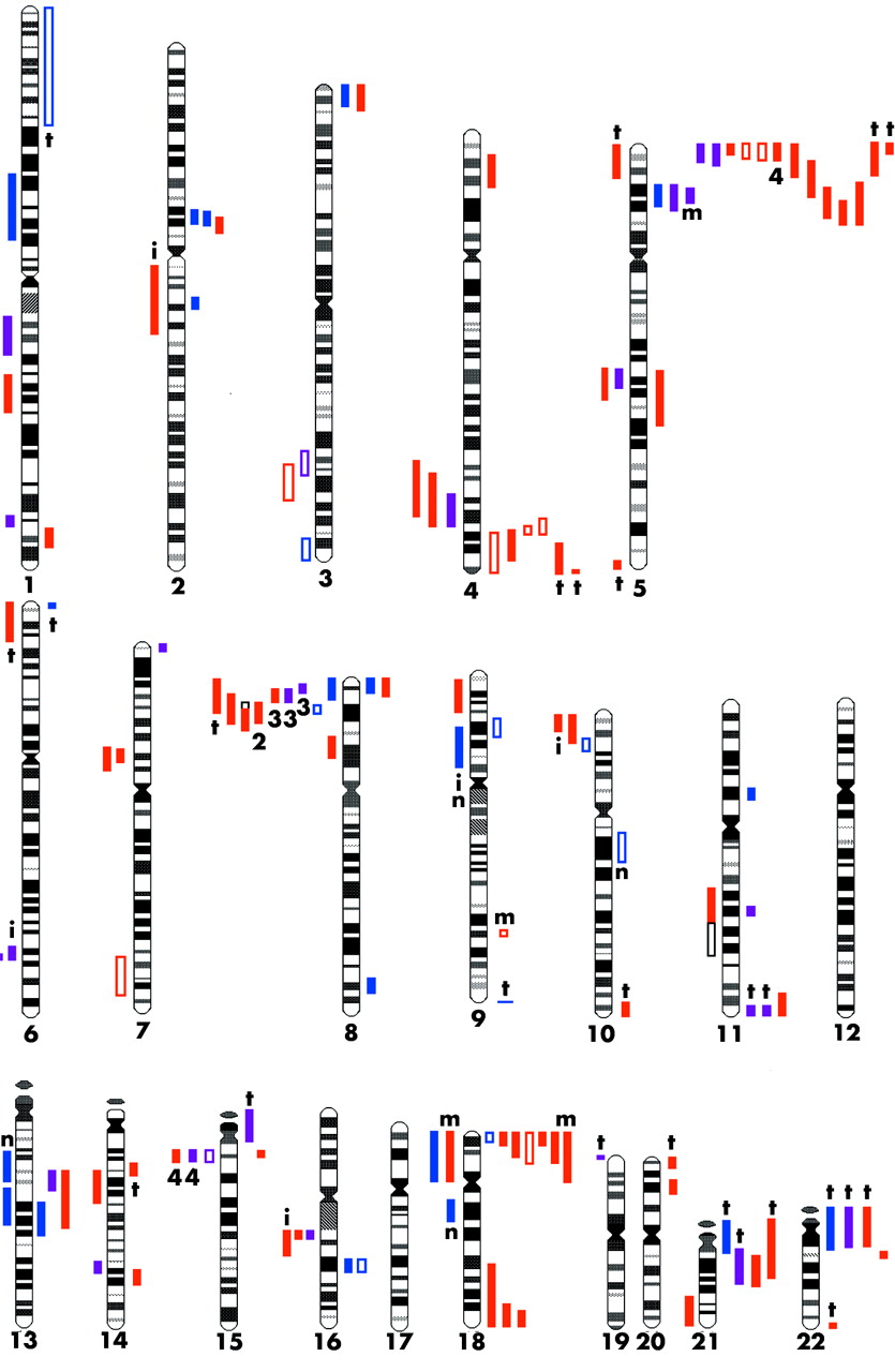
Idiograms with extent of duplications on the left hand side and deletions on the right hand side. Group 1 imbalances are in blue, group 2 in purple, and group 3 in red. Filled coloured bars are UBCAs from peer reviewed papers; open coloured boxes are from abstracts only. Open black boxes indicate alternative interpretations according to the authors concerned. Figures in black give the number of times independent families with the same rearrangement have been reported (for example, four times). t, translocation; i, insertion; m, mosaicism in a parent; n, the four exceptional UBCAs that were not directly transmitted.

Idiograms with the position at which EVs occur marked by arrows. Group 1 EV imbalances are in blue; group 2 EV in purple, and group 3 EV in red. Figures give the number of times independent families with the same rearrangement have been reported (for example, eight times).
Acknowledgments
P Jacobs, A Sharp, and N Cross are thanked for their helpful comments on this review. VMaloney is thanked for constructing the idiograms and J Gladding for her help with the preparation of the manuscript.
REFERENCES
- ↵
- ↵
- ↵
- ↵
- ↵
- ↵
- ↵
- ↵
- ↵
- ↵
- ↵
- ↵
- ↵
- ↵
- ↵
- ↵
- ↵
- ↵
- ↵
- ↵
- ↵
- ↵
- ↵
- ↵
- ↵
- ↵
- ↵
- ↵
- ↵
- ↵
- ↵
- ↵
- ↵
- ↵
- ↵
- ↵
- ↵
- ↵
- ↵
- ↵
- ↵
- ↵
- ↵
-
- ↵
- ↵
- ↵
- ↵
- ↵
- ↵
- ↵
- ↵
- ↵
- ↵
- ↵
- ↵
- ↵
-
- ↵
- ↵
- ↵
- ↵
- ↵
- ↵
- ↵
- ↵
- ↵
- ↵
- ↵
- ↵
- ↵
- ↵
- ↵
- ↵
- ↵
- ↵
- ↵
- ↵
- ↵
- ↵
- ↵
- ↵
- ↵
- ↵
- ↵
- ↵
-
- ↵
- ↵
- ↵
- ↵
- ↵
- ↵
- ↵
- ↵
- ↵
- ↵
- ↵
- ↵
- ↵
- ↵
- ↵
- ↵
- ↵
- ↵
- ↵
- ↵
- ↵
- ↵
- ↵
- ↵
- ↵
- ↵
- ↵
- ↵
- ↵
- ↵
- ↵
- ↵
- ↵
- ↵
- ↵
- ↵
- ↵
- ↵
- ↵
- ↵
- ↵
- ↵
- ↵
- ↵
- ↵
- ↵
- ↵
- ↵
- ↵
- ↵
- ↵
- ↵
- ↵
- ↵
- ↵
- ↵
- ↵
- ↵
- ↵
- ↵
- ↵
- ↵
- ↵
- ↵
- ↵
- ↵
- ↵
- ↵
- ↵
- ↵
- ↵
- ↵
- ↵
- ↵
- ↵
- ↵
- ↵
- ↵
- ↵
- ↵
Footnotes
-
Competing interests: none declared